BIO 004 · Human Anatomy
Blood Vessels, Structure & Types
Block 3 · Module 4: Blood Vessels, Structure and Types
A reference for the blood vessels video and lab. This page covers the three tunics of a vessel wall, the five vessel types, the kinds of artery, capillary, and vein, and the circulatory routes blood follows.
How to use this sheet Toggle the toolbar above. Notes prints the full reference for review. Study prints as a fill-in-the-blank worksheet. Print it, then write each definition while you watch the video or read your book. Quiz me is on-screen typing practice: type the term, click Reveal to check yourself. The comparison grids respond to Study and Quiz too, with a Reveal button on each row.
The Foundations video gives you a complete foundational understanding of this topic, enough on its own for a foundational course. Learn it first, then move on to the Deep dive, which adds the majors-level material: the capillary types, the venous return structures, and the circulatory routes.
- Name the three tunics of a vessel wall and what each contains.
- Compare the five vessel types from artery to vein.
- Distinguish the kinds of artery, capillary, and vein.
- Describe the circulatory routes, including portal systems and anastomoses.
Your pre-work
Work through these the evening before class. None of it is turned in. It is how you learn the material and build your spaced recall.
This is more than a checklist. Ticking these boxes is the start, not the finish. Committing this material to memory and being able to apply it takes considerable time and repeated effort. You are not done when the boxes are checked. Put in the real hours, and keep coming back for frequent recall and review until the material is genuinely yours.
The three tunics
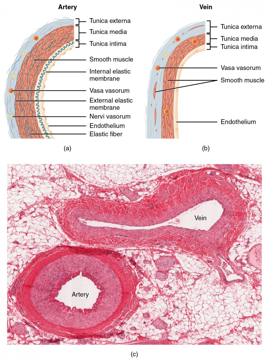
Add a labeled cross-section of an artery and a vein showing the tunica intima, media, and externa.
The vessel types in series
Add a labeled diagram from artery to arteriole to capillary to venule to vein.
The capillary types
Add labeled views of a continuous capillary, a fenestrated capillary, and a sinusoid.
Blood Vessels, an Overview
Blood vessels are the body's plumbing. Blood leaves the heart in arteries, reaches the tissues through capillaries, and returns in veins.
- Blood vessela tube that carries blood through the body
- Arterya vessel that carries blood away from the heart
- Veina vessel that carries blood toward the heart
- Capillarythe smallest vessel, where exchange between the blood and the tissues takes place
- Lumenthe hollow interior of a vessel, the channel the blood flows through
- The vascular sequencearteries branch into arterioles, then capillaries, which merge into venules, then veins
The Three Tunics
The wall of an artery or a vein is built in three concentric layers, the tunics. A capillary wall is just the innermost layer. Compare the three.
| Tunic | Position | Tissue | Note |
|---|---|---|---|
| Tunica intima | the innermost layer | a lining of simple squamous endothelium on a basement membrane | the only layer a capillary has |
| Tunica media | the middle layer | smooth muscle and elastic fibers | the thickest layer in arteries; its smooth muscle sets the vessel's diameter |
| Tunica externa | the outermost layer | mostly collagen fibers | anchors the vessel to the surrounding tissue |
The Vessel Types
Blood passes through five kinds of vessel in series, each with a wall built for its job. Compare them.
| Vessel | Direction of flow | Wall | Role |
|---|---|---|---|
| Arteries | carry blood away from the heart | thick walls with a heavy tunica media | carry blood under high pressure |
| Arterioles | away from the heart, the smallest arteries | thinner walls, mostly smooth muscle | adjust how much blood enters a capillary bed |
| Capillaries | connect arterioles to venules | a single layer, the tunica intima only | the exchange vessels; gases and nutrients cross here |
| Venules | toward the heart, the smallest veins | thin walls | collect blood drained from the capillaries |
| Veins | carry blood toward the heart | thinner walls and a wide lumen, with valves | carry blood under low pressure |
Arteries
Arteries carry blood away from the heart under high pressure. They come in three sizes, and the smallest control how much blood reaches the tissues.
- Elastic arteriesthe largest arteries, such as the aorta; their walls are rich in elastic fibers that stretch with each heartbeat and recoil between beats
- Muscular arteriesmedium-sized arteries that deliver blood to specific organs; their tunica media is mostly smooth muscle
- Arteriolesthe smallest arteries, the gatekeepers that control blood flow into the capillary beds
- Vasoconstrictionthe narrowing of a vessel as the smooth muscle of its wall contracts
- Vasodilationthe widening of a vessel as the smooth muscle of its wall relaxes
Capillaries
Capillaries are the only vessels thin enough for exchange. They come in three types, leakier as the tissue's job demands. Compare them.
| Capillary type | Wall | Where found |
|---|---|---|
| Continuous capillary | an unbroken endothelial lining | the most common type, in muscle, skin, and the lungs |
| Fenestrated capillary | an endothelium with small pores, called fenestrations | where rapid absorption or filtration happens, as in the kidney and small intestine |
| Sinusoid | a wide, leaky vessel with large gaps between the cells | where even whole cells must cross, as in the liver, spleen, and bone marrow |
- Capillary beda network of capillaries supplied by a single arteriole
- Precapillary sphinctera ring of smooth muscle that opens or closes the entrance to a capillary
- Vascular shunta direct channel that lets blood bypass the capillaries of a bed when exchange is not needed
Veins
Veins return blood to the heart under low pressure. Because the pressure is low, they rely on valves and on nearby muscles to keep blood moving the right way.
- Venulethe smallest vein, collecting blood as it leaves a capillary bed
- Veina vessel returning blood to the heart, with a thinner wall and a wider lumen than the matching artery
- Venous valvesflaps of the tunica intima, common in the limb veins, that keep blood from flowing backward
- Skeletal muscle pumpcontracting limb muscles that squeeze the deep veins and push blood toward the heart
- Blood reservoirsthe veins as a group, which at rest hold the largest share of the body's blood
Circulatory Routes
Most blood follows the simple loop of artery to capillary to vein, but a few routes are arranged differently.
- Pulmonary circuitthe route from the right side of the heart to the lungs and back to the left side
- Systemic circuitthe route from the left side of the heart out to the body and back to the right side
- Portal systeman arrangement in which blood passes through two capillary beds in series before returning to the heart, as in the hepatic portal system that routes blood through the liver
- Anastomosisa place where two vessels join, giving blood an alternate route if one path is blocked
- Arteriovenous anastomosisa direct connection from an artery to a vein that bypasses the capillaries
See also: The Heart, the pump that drives these vessels, and Blood Vessel Disorders and Fetal Circulation, the next page in this block.
Study questions
Work on answering these in writing, in your own words. They are the questions to bring to class, and good practice for the reasoning the exams ask for.
- Compare the three vessel types, artery, capillary, and vein, by wall thickness and the job each does.
- Why do veins have valves and arteries do not? Tie your answer to blood pressure.
- Trace blood from the left ventricle to a capillary bed and back to the right atrium, naming the vessel types in order.
- Explain how the structure of a capillary suits exchange between blood and tissue.
Step 2 . Retrieval check
Now explain it back, in your own words.
In 60 words or more, pull together what the video just taught you. Include the key concepts. This is the point where the learning actually sticks. After you submit, your spaced-recall cards for this topic unlock.