BIO 004 · Human Anatomy
Anatomy of the Cell
Cell Anatomy
A reference for the cell anatomy video and lab. The cell is the smallest living unit of the body. Every tissue, organ, and system is built from cells, so the structures named here are the foundation for the histology page that follows.
How to use this sheet
Notes prints the full reference for review.
Study prints as a fill-in-the-blank worksheet to complete while you watch the video or read.
Quiz me is on-screen practice: type the term, or click Reveal to check yourself.
The tables and sequences respond to Study and Quiz too, with a Reveal button on each row.
- Name the three basic regions of a generalized cell and state what each one contains.
- Describe the plasma membrane, distinguish integral from peripheral proteins, and name its surface specializations.
- Identify the membranous and non-membranous organelles and state the job of each.
- Explain how the nucleus stores the genetic material and how the nuclear pores control its traffic.
- Trace the path a product follows through the endomembrane system, from the ER to the cell surface.
Your pre-work
Work through these the evening before class. None of it is turned in. It is how you learn the material and build your spaced recall.
This is more than a checklist. Ticking these boxes is the start, not the finish. Committing this material to memory and being able to apply it takes considerable time and repeated effort. You are not done when the boxes are checked. Put in the real hours, and keep coming back for frequent recall and review until the material is genuinely yours.
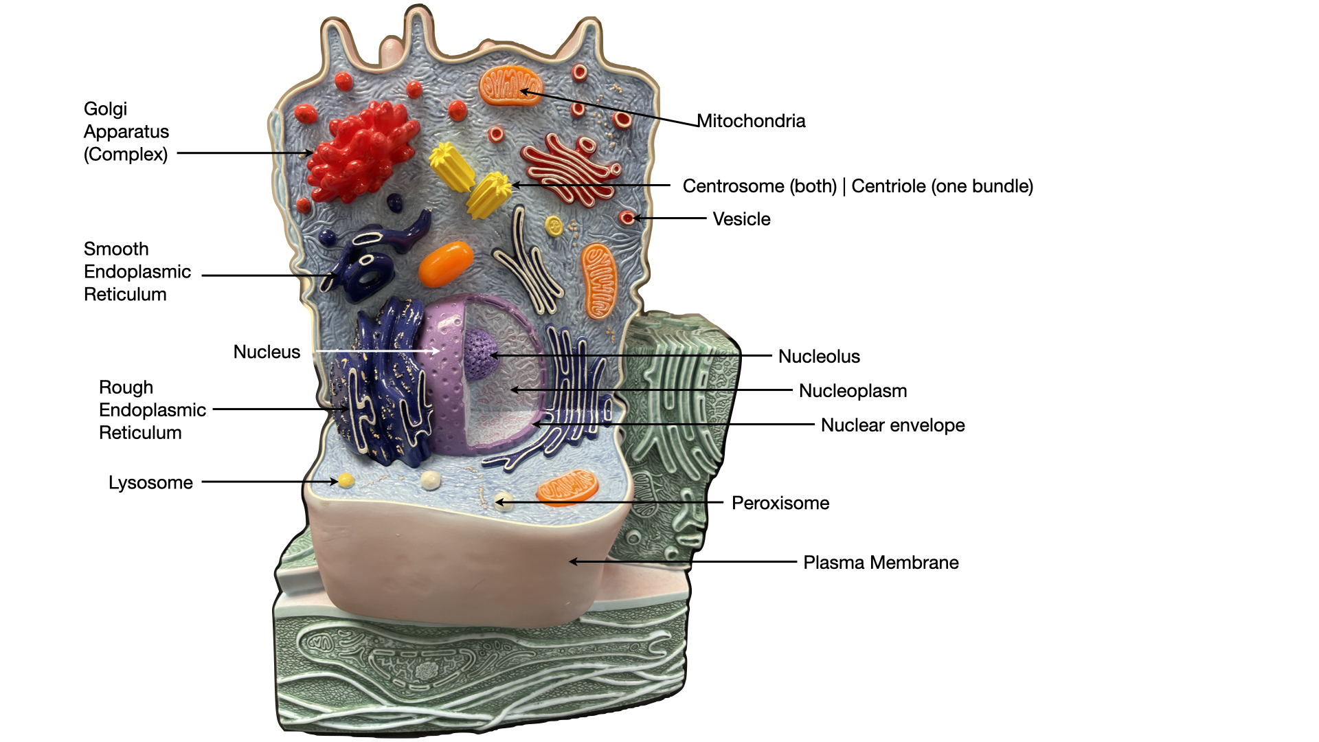
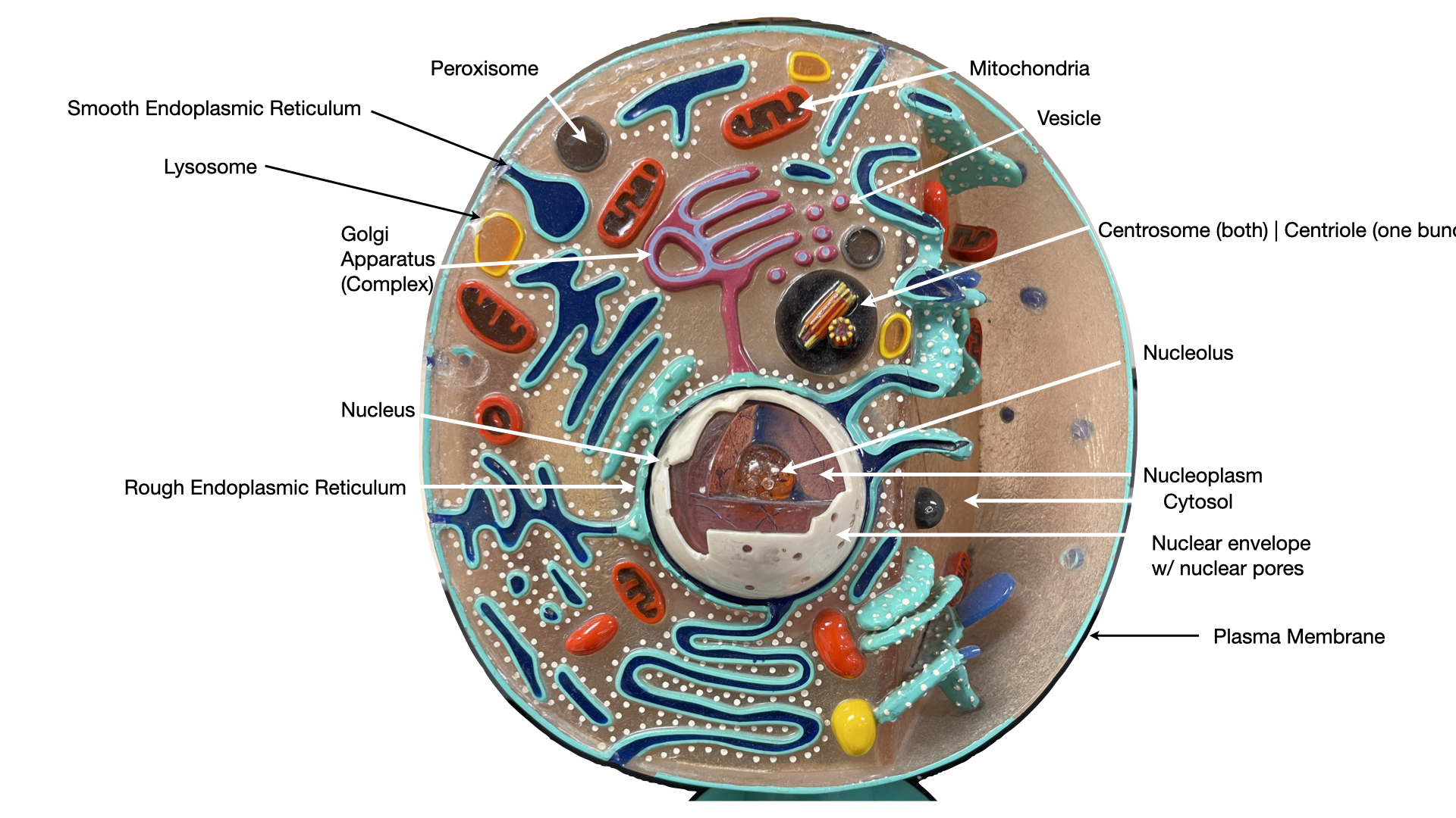
The Generalized Cell
Cells vary widely, but a composite model shows the three regions and the structures common to most body cells.
- Generalized cella composite model showing the features shared by most body cells
- Plasma membranethe outer boundary that encloses the cell and separates it from its surroundings
- Cytoplasmeverything between the plasma membrane and the nucleus, the cytosol plus the organelles
- Nucleusthe control center, usually the largest structure in the cell
The Plasma Membrane
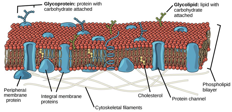
The outer boundary of the cell, and a selective barrier that controls which substances enter and leave. Compare its components by where each one sits and what each one does.
Membrane structure
| Component | Location in the membrane | Role |
|---|---|---|
| Phospholipid bilayer | a double layer of phospholipids, the basic fabric of the membrane | forms the continuous sheet; the fluid mosaic model describes proteins drifting within it |
| Integral proteins | firmly embedded in the bilayer; most span it completely as transmembrane proteins | form the channels, carriers, and receptors |
| Peripheral proteins | attached loosely to one face of the membrane or to integral proteins, not embedded | act as enzymes and brace the cell shape |
| Cholesterol | wedged among the phospholipids | stabilizes the membrane and keeps it fluid |
| Glycocalyx | the fuzzy external coat of glycoproteins and glycolipids | the cell's identity badge, used in recognition and adhesion |
Surface specializations
| Specialization | Description | Function |
|---|---|---|
| Microvilli | finger-like extensions of the cell surface | increase surface area for absorption |
| Cilia | short hair-like extensions, built on a 9+2 microtubule core (nine doublets around a central pair) | sweep substances across the cell surface |
| Flagellum | a single long extension, also a 9+2 microtubule core | propels the cell; the only human example is the sperm tail |
The Nucleus
The control center of the cell. It stores the genetic code and directs the building of proteins.
- Nuclear envelopethe double membrane that encloses the nucleus, its outer membrane is continuous with the rough ER
- Nuclear poresprotein-lined channels through the envelope that regulate traffic both ways, messenger RNA and ribosomal subunits move out while proteins needed inside move in
- Nucleoplasmthe fluid inside the nucleus that suspends the other nuclear structures
- Nucleolusa dense body inside the nucleus where ribosomal subunits are assembled
- Chromatinthe loose, threadlike form of DNA wound on proteins, present when the cell is not dividing
- Chromosomesthe tightly coiled form of chromatin, visible when the cell divides
Cytoplasm and Organelles
The cytoplasm is the cytosol, a gel-like fluid that suspends the organelles, plus the organelles themselves, the small structures that each carry out a specific job. Compare them by structure and function.
Membranous organelles
| Organelle | Structure | Function |
|---|---|---|
| Rough ER | a ribosome-studded membrane network | makes and packages proteins |
| Smooth ER | a tubular membrane network with no ribosomes | makes lipids and stores calcium |
| Golgi apparatus | a stack of flattened membrane sacs | sorts, packages, and ships cell products |
| Lysosomes | membrane sacs of digestive enzymes | break down worn parts and debris |
| Peroxisomes | membrane sacs of enzymes | neutralize toxins and free radicals |
| Mitochondria | double-membraned organelles | produce most of the cell's ATP |
| Vesicles | small membrane sacs | move substances within the cell |
Non-membranous organelles
| Organelle | Structure | Function |
|---|---|---|
| Ribosomes | tiny particles of RNA and protein, free in the cytosol or attached to the rough ER | build proteins |
| Cytoskeleton | an internal protein framework | gives the cell shape and allows movement |
| Microfilaments | the thinnest fibers, made of actin | support the cell surface and enable movement |
| Intermediate filaments | rope-like fibers | resist pulling forces and give the cell strength |
| Microtubules | the thickest fibers, hollow tubes | form tracks for transport inside the cell |
| Centrosome | a region near the nucleus | organizes the microtubules |
| Centrioles | paired rod-shaped structures within the centrosome, nine microtubule triplets in a 9+0 array | organize the spindle during cell division |
The Path Materials Take Through the Cell
The membranous organelles connect into one route, the endomembrane system: the nuclear envelope, the ER, the Golgi apparatus, vesicles, and lysosomes. The numbered path below traces a secreted protein in order, from where it is made to where it leaves the cell. The mechanism belongs to physiology and is not covered here.
- Rough ERthe start of the path, where a new protein is made and enters the endomembrane system
- Transport vesiclebuds from the ER and carries the product to the Golgi apparatus
- Golgi apparatusthe mid-path stop, where the product is sorted, modified, and packaged
- Secretory vesiclethe carrier on the final leg, from the Golgi to the plasma membrane or to a lysosome
- Exocytosisthe exit point, where the vesicle fuses with the plasma membrane and the product leaves the cell
Supporting structures on the route
- Endocytosisthe reverse direction, where material is taken into the cell inside a newly formed vesicle
- Microtubulesthe tracks the vesicles follow between stops on the path
- Nuclear poresthe gateway on the path between the nucleus and the cytoplasm, where messenger RNA exits
Cells Vary With Their Job
No single cell looks like the generalized model. A cell's shape and its mix of organelles match the work it does: structure follows job, and a cell built for a task carries the organelles that task needs. Compare three examples.
| Cell type | Structural adaptation | Why it fits the job |
|---|---|---|
| Muscle cell | long and packed with mitochondria | contraction demands a constant, heavy supply of ATP |
| Red blood cell | loses its nucleus and organelles | frees interior space for oxygen-carrying contents |
| White blood cell | changes shape freely | can squeeze between cells to reach a site of infection |
See also: Histology: The Four Tissue Types, the companion page in this module, where cells are organized into tissues.
Build the word
Term breakdown
Organelle names describe what the part looks like or what it does. Learn the roots and the names stop being arbitrary.
- cyt- / -cytecellcytoplasm, cytosol, cytoskeleton
- plasmaformed, moldedplasma membrane, cytoplasm
- nucle-kernelnucleus, nucleolus
- mito- + chondr-thread + granulemitochondrion
- endo-withinendoplasmic reticulum, endosome
- reticul-little netendoplasmic reticulum
- lys-to split, loosenlysosome
- -somebodyribosome, lysosome, peroxisome
- micro-smallmicrovilli, microtubule, microfilament
- vill-shaggy hairmicrovilli
- cili-eyelashcilia
- flagell-whipflagellum
- centr-centercentriole, centrosome
- phag- / pino-eat / drinkphagocytosis, pinocytosis
- -plasmformed substancecytoplasm, nucleoplasm
Draw it from memory Sketch and label before you check
Work on blank paper, from memory, before you look back at the figures. Drawing the cell forces you to place each organelle, not just name it.
- Draw a generalized cell. Label the plasma membrane, nucleus, nucleolus, and at least six organelles in the cytoplasm.
- Draw the plasma membrane in cross-section. Label the phospholipid bilayer (heads and tails), an integral (transmembrane) protein, and a peripheral protein.
- Draw the endomembrane path. From memory, trace a protein from the rough ER to a transport vesicle, to the Golgi apparatus, to a secretory vesicle, to the plasma membrane. Label every station.
Then check yourself. Compare your drawing against the figures and tables above. Whatever you left out or placed wrong is tonight's study list. Redraw it until it comes out right without looking.
Study questions
Work on answering these in writing, in your own words. They are the questions to bring to class, and good practice for the reasoning the exams ask for.
- Name the three regions of a generalized cell and state what each one contains.
- Trace a secreted protein through the endomembrane system, from where it is made to where it leaves the cell.
- Compare membranous and non-membranous organelles and give two examples of each.
- Explain how the structure of the plasma membrane makes it a selective barrier.
Step 2 . Retrieval check
Now explain it back, in your own words.
In 60 words or more, pull together what the video just taught you. Include the key concepts. This is the point where the learning actually sticks. After you submit, your spaced-recall cards for this topic unlock.