BIO 004 · Human Anatomy · Week 4
The Lymphatic System
Drainage, transport, and defense. The vessels that reclaim fluid, the cells that police it, and the organs that house them.
Overview
What the lymphatic system does
Three jobs
- Fluid balance: reclaims interstitial fluid that leaks from blood capillaries and returns it to the blood.
- Fat absorption: lacteals in the intestinal villi absorb dietary fats.
- Immune defense: houses and transports the lymphocytes that screen fluid for pathogens.

Dr. Rennie, lecture figure
Vessels
Lymph and lymphatic capillaries
Where lymph starts
- Lymphatic capillaries are blind-ended, more permeable than blood capillaries; overlapping endothelial minivalves let fluid in but not out.
- Found almost everywhere except avascular tissue, bone, teeth, bone marrow, and the CNS.
- Lacteals: the lymphatic capillaries in the center of each intestinal villus that absorb fat (chyle).

Dr. Rennie, lecture figure
Vessels
Collecting vessels, trunks, and ducts
From capillary to bloodstream
- Collecting vessels have three tunics and many valves; travel as superficial and deep sets.
- They drain into nine trunks, then into two ducts.
- Thoracic duct drains three-quarters of the body (everything below the diaphragm plus the upper left); right lymphatic duct drains the upper right.
- The ducts empty into the subclavian veins.

Dr. Rennie, lecture figure
Vessels
How lymph moves
- No central pump: lymph is pushed by the skeletal muscle pump as muscles squeeze the vessels.
- The respiratory pump moves it with pressure changes during breathing.
- Smooth muscle in vessel walls contracts, and valves keep lymph moving one way.
Cells
The Lymphocytes
Three cell lines defend the body, made in the marrow and matured in the primary organs.
Cells
B cells, T cells, and NK cells
Three types
- T lymphocytes: about 80% of circulating lymphocytes; cell-mediated immunity; mature in the thymus.
- B lymphocytes: about 10 to 15%; humoral (antibody) immunity; mature in the red bone marrow.
- NK (natural killer) cells: attack foreign cells, virus-infected cells, and cancer cells.

Dr. Rennie, lecture figure
Cells
Lymphopoiesis and the primary organs
Made in the marrow, schooled in the primaries
- Lymphopoiesis: lymphocytes arise from stem cells in the red bone marrow.
- Primary lymphatic organs (red bone marrow and thymus) are where lymphocytes mature and become immunocompetent.
- Secondary lymphatic organs (lymph nodes, spleen, tonsils, MALT) are where mature lymphocytes encounter antigens.

Dr. Rennie, lecture figure
Tissue
Lymphatic Tissue & Follicles
Reticular connective tissue packed with lymphocytes, organized into follicles.
Tissue
Lymphatic tissue and follicles
The building block
- All lymphatic tissues and organs (except the thymus) are built on reticular connective tissue.
- Lymphocytes are the dominant leukocyte.
- Follicles (nodules): dense spherical clusters with a paler germinal center dominated by dividing B cells.
- Aggregated follicles: Peyer’s patches in the ileum and the tonsils in the pharynx.

Dr. Rennie, lecture figure
Organs
Tonsils
The pharyngeal ring
- Tonsils are follicle clusters in the walls of the pharynx, positioned to catch pathogens in inhaled air and swallowed food.
- Deep invaginations called crypts trap material for the lymphocytes to sample.
- Tonsillitis: inflammation and swelling of the tonsils, usually from infection.

Dr. Rennie, lecture figure
Organs
Lymph nodes
The filters
- Functions: filter lymph and activate the immune response; fixed macrophages phagocytose debris and pathogens.
- Flow through a node: lymph enters by several afferent vessels, percolates through sinuses, and leaves by fewer efferent vessels at the hilum, so it lingers to be cleaned.
- Clustered in the cervical, axillary, and inguinal regions.

Dr. Rennie, lecture figure
Organs
Thymus
Where T cells are schooled
- Larger and more active in children; involutes with age.
- Cortex: rapid T-cell division and selection. Medulla: maturing T cells and thymic (Hassall’s) corpuscles.
- Built on epithelial reticular cells (not reticular CT), which secrete hormones that drive T-cell maturation.
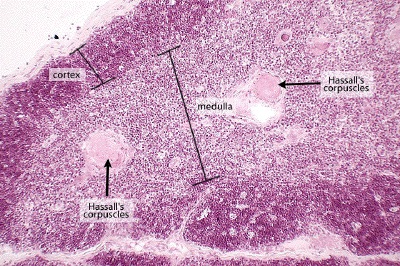
Thymus, cortex, medulla, and Hassall corpuscles
Organs
Spleen
The largest lymphatic organ
- Largest collection of lymphatic tissue; filters blood rather than lymph.
- White pulp: lymphocytes around central arteries, the immune function. Red pulp: sinuses and cords that remove old red blood cells and store platelets.
- Blood flow: splenic artery to white and red pulp to venous sinuses to splenic vein.

Dr. Rennie, lecture figure
Clinical
Spleen and lymphatic disorders
- Mononucleosis or leukemia can enlarge the spleen (splenomegaly), stretching its capsule so it ruptures easily, a dangerous internal bleed.
- We can live without the spleen because the liver and bone marrow take over its filtering; the trade-off after splenectomy is higher infection risk.
- Elephantiasis: parasitic blockage of lymphatic vessels causes massive limb swelling; treated by managing the infection and the swelling.
- Blocked drainage anywhere causes lymphedema.
Wrap-up
Key takeaways
- The lymphatic system reclaims fluid, absorbs fat at the lacteals, and defends the body.
- Lymph flows one way: capillaries to collecting vessels to nine trunks to two ducts to the subclavian veins.
- Three lymphocytes: T (thymus, cell-mediated), B (marrow, antibodies), NK.
- Primary organs (marrow, thymus) make and mature cells; secondary organs (nodes, spleen, tonsils) deploy them.
- Nodes filter lymph; the spleen filters blood (white pulp defends, red pulp recycles).
Keep studying
Take it with you
Review the lecture
These slides

Open the deck on your phone.
Practice from memory
Study-question guide

Questions only.