Digestive Anatomy Human Anatomy-Dr. Rennie
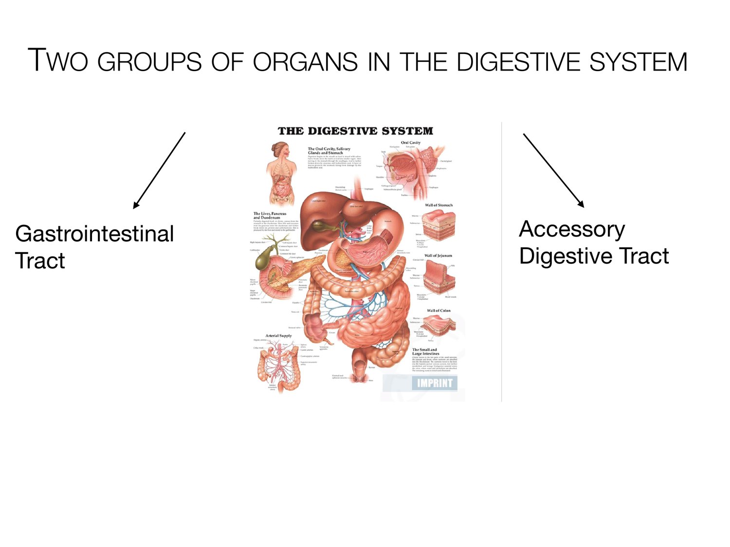
TWO GROUPS OF ORGANS IN THE DIGESTIVE SYSTEM Gastrointestinal Accessory Tract Digestive Tract
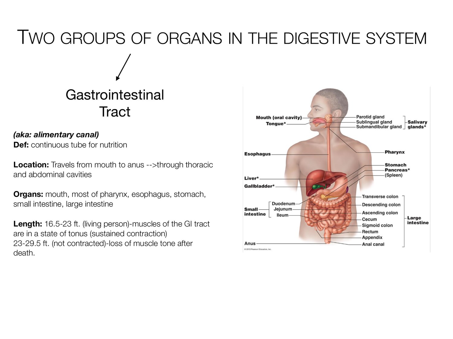
TWO GROUPS OF ORGANS IN THE DIGESTIVE SYSTEM Gastrointestinal Tract (aka: alimentary canal) Def: continuous tube for nutrition Location: Travels from mouth to anus -->through thoracic and abdominal cavities Organs: mouth, most of pharynx, esophagus, stomach, small intestine, large intestine Length: 16.5-23 ft. (living person)-muscles of the GI tract are in a state of tonus (sustained contraction) 23-29.5 ft. (not contracted)-loss of muscle tone after death.
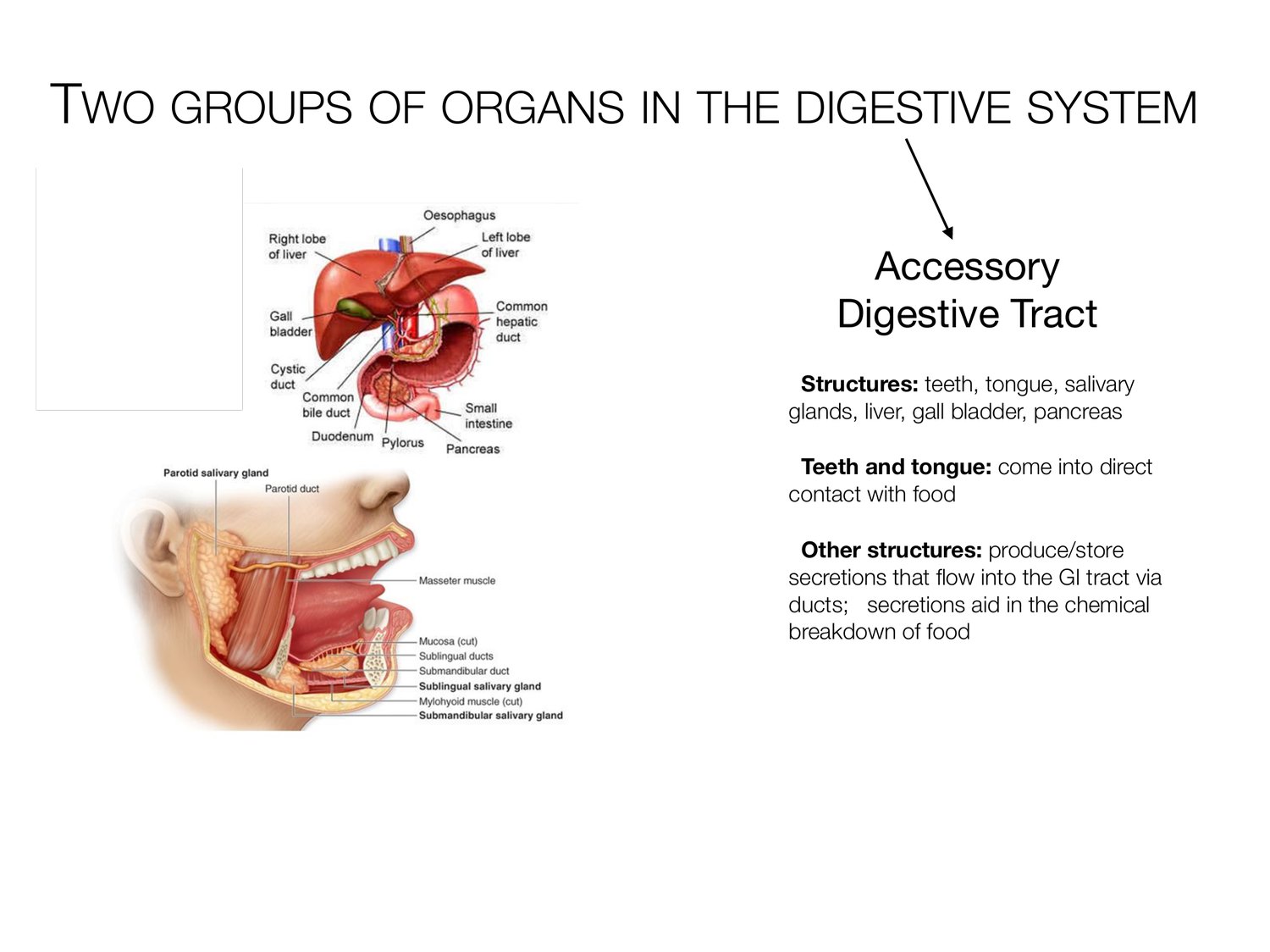
TWO GROUPS OF ORGANS IN THE DIGESTIVE SYSTEM Accessory Digestive Tract Structures: teeth, tongue, salivary glands, liver, gall bladder, pancreas Teeth and tongue: come into direct contact with food Other structures: produce/store secretions that ow into the GI tract via ducts; secretions aid in the chemical breakdown of food fl
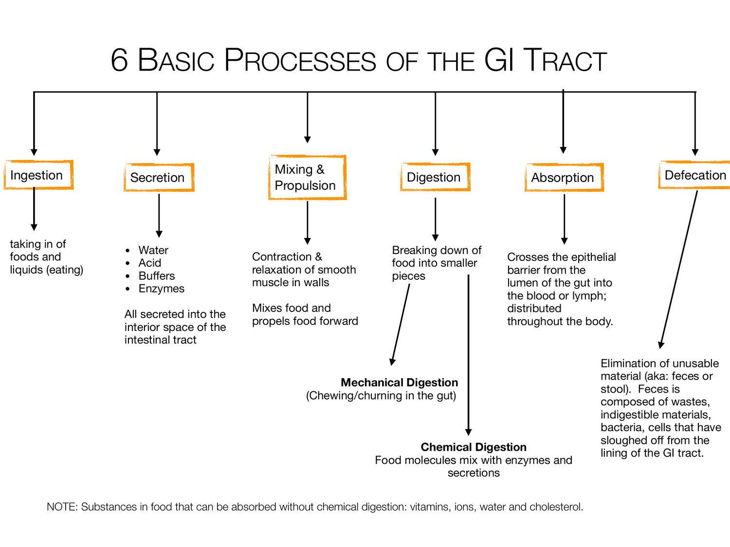
6 BASIC PROCESSES OF THE GI TRACT Ingestion Mixing & Defecation Secretion Digestion Absorption Propulsion taking in of foods and • Water Contraction & Breaking down of Crosses the epithelial liquids (eating) • Acid relaxation of smooth food into smaller barrier from the • Bu ers muscle in walls pieces lumen of the gut into • Enzymes the blood or lymph; Mixes food and distributed All secreted into the propels food forward throughout the body. interior space of the intestinal tract Elimination of unusable material (aka: feces or Mechanical Digestion stool). Feces is (Chewing/churning in the gut) composed of wastes, indigestible materials, bacteria, cells that have sloughed o from the Chemical Digestion lining of the GI tract. Food molecules mix with enzymes and secretions NOTE: Substances in food that can be absorbed without chemical digestion: vitamins, ions, water and cholesterol. ff ff
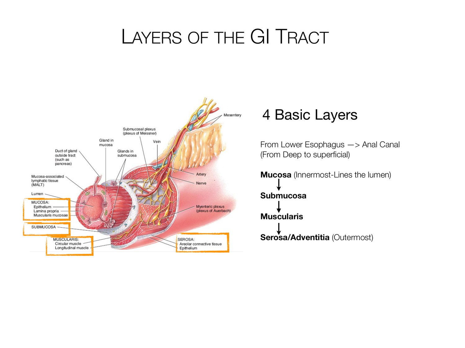
LAYERS OF THE GI TRACT 4 Basic Layers From Lower Esophagus -> Anal Canal (From Deep to super cial) Mucosa (Innermost-Lines the lumen) Submucosa Muscularis Serosa/Adventitia (Outermost) fi
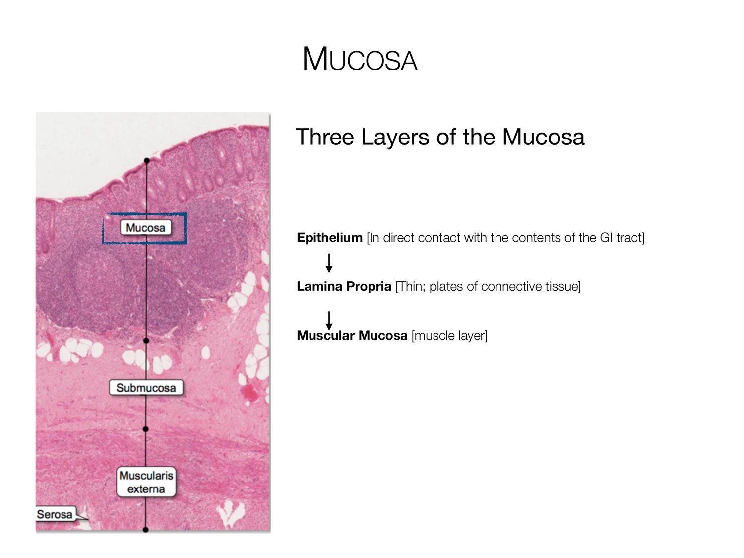
MUCOSA Three Layers of the Mucosa Epithelium [In direct contact with the contents of the GI tract] Lamina Propria [Thin; plates of connective tissue] Muscular Mucosa [muscle layer]
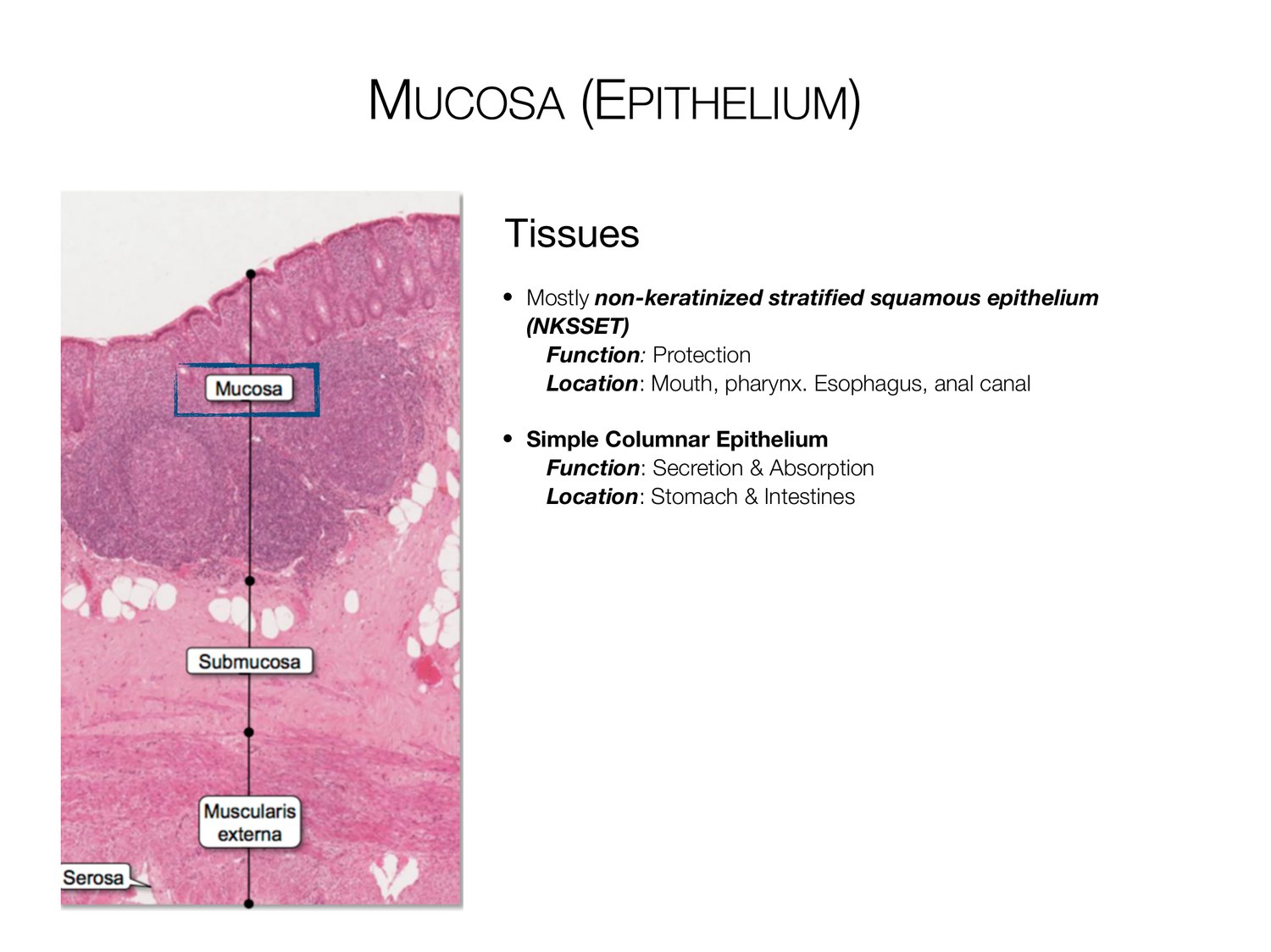
MUCOSA (EPITHELIUM) Tissues • Mostly non-keratinized strati ed squamous epithelium (NKSSET) Function: Protection Location: Mouth, pharynx. Esophagus, anal canal • Simple Columnar Epithelium Function: Secretion & Absorption Location: Stomach & Intestines fi
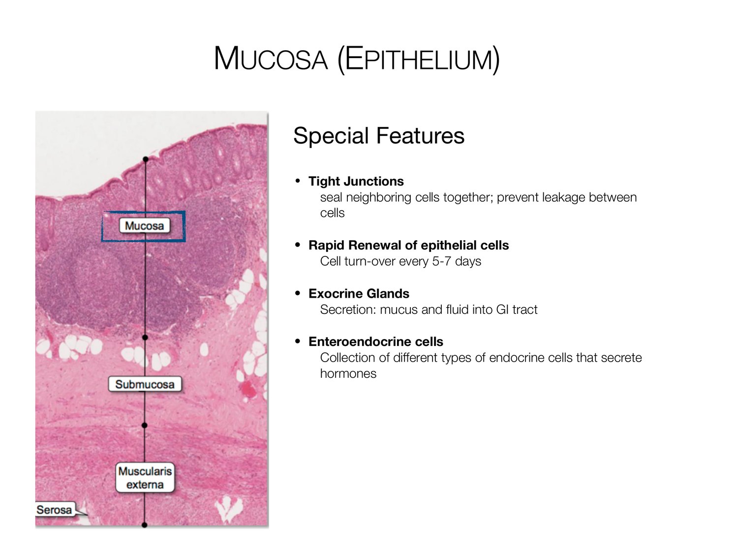
MUCOSA (EPITHELIUM) Special Features • Tight Junctions seal neighboring cells together; prevent leakage between cells • Rapid Renewal of epithelial cells Cell turn-over every 5-7 days • Exocrine Glands Secretion: mucus and uid into GI tract • Enteroendocrine cells Collection of different types of endocrine cells that secrete hormones fl
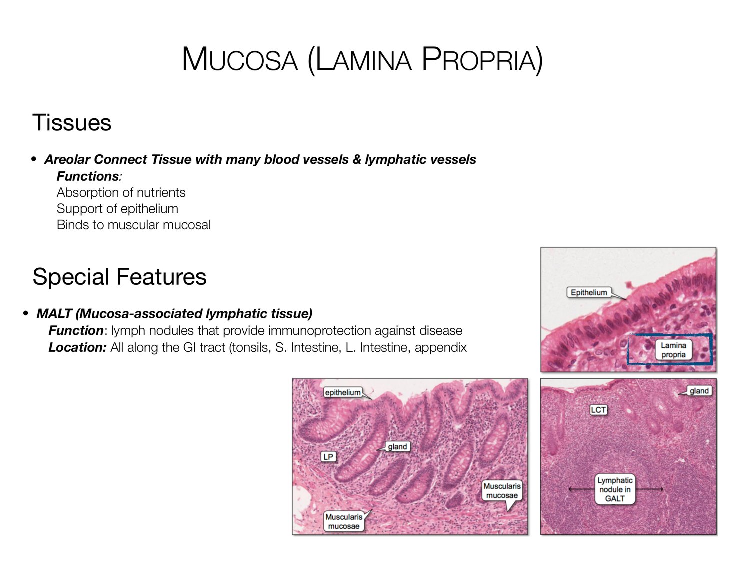
MUCOSA (LAMINA PROPRIA) Tissues • Areolar Connect Tissue with many blood vessels & lymphatic vessels Functions: Absorption of nutrients Support of epithelium Binds to muscular mucosal Special Features • MALT (Mucosa-associated lymphatic tissue) Function: lymph nodules that provide immunoprotection against disease Location: All along the GI tract (tonsils, S. Intestine, L. Intestine, appendix
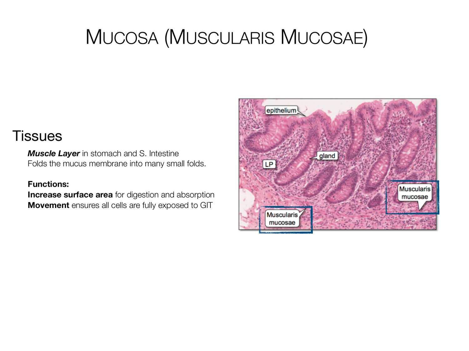
MUCOSA (MUSCULARIS MUCOSAE) Tissues Muscle Layer in stomach and S. Intestine Folds the mucus membrane into many small folds. Functions: Increase surface area for digestion and absorption Movement ensures all cells are fully exposed to GIT
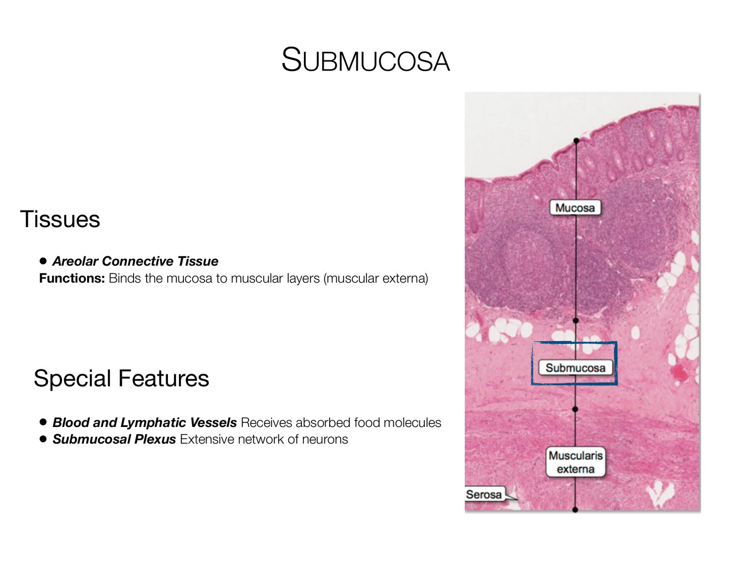
SUBMUCOSA Tissues • Areolar Connective Tissue Functions: Binds the mucosa to muscular layers (muscular externa) Special Features • Blood and Lymphatic Vessels Receives absorbed food molecules • Submucosal Plexus Extensive network of neurons
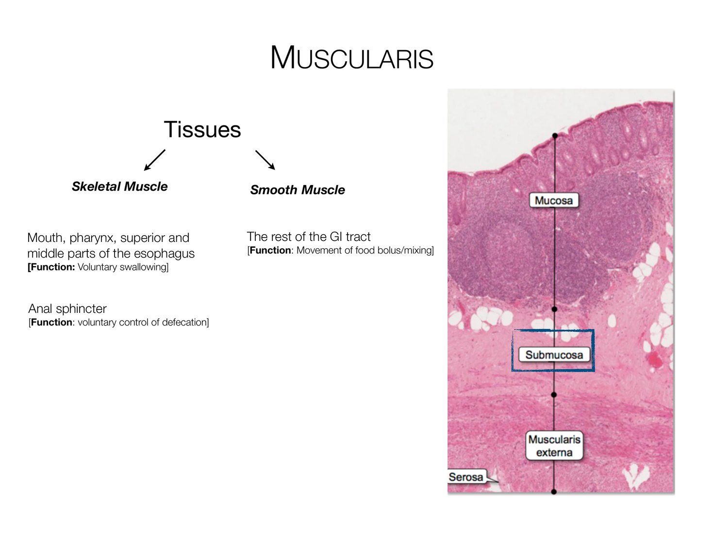
MUSCULARIS Tissues Skeletal Muscle Smooth Muscle Mouth, pharynx, superior and The rest of the GI tract middle parts of the esophagus [Function: Movement of food bolus/mixing] [Function: Voluntary swallowing] Anal sphincter [Function: voluntary control of defecation]
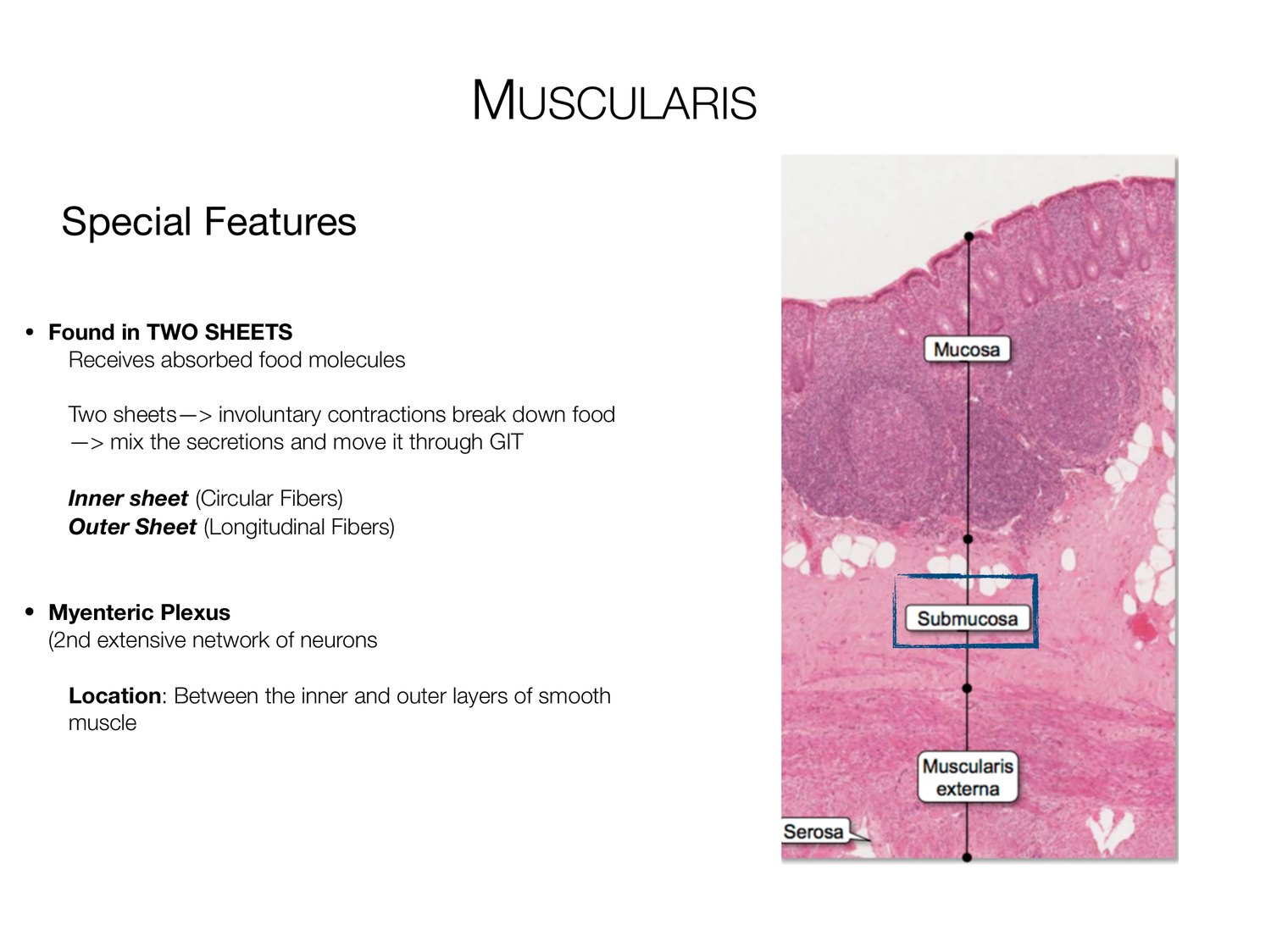
MUSCULARIS Special Features • Found in TWO SHEETS Receives absorbed food molecules Two sheets-> involuntary contractions break down food -> mix the secretions and move it through GIT Inner sheet (Circular Fibers) Outer Sheet (Longitudinal Fibers) • Myenteric Plexus (2nd extensive network of neurons Location: Between the inner and outer layers of smooth muscle
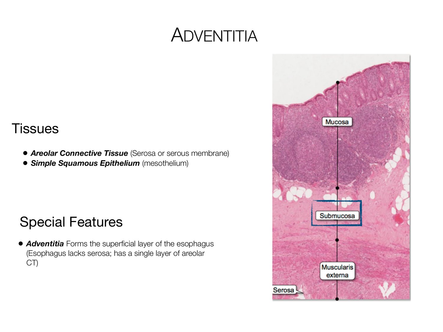
ADVENTITIA Tissues • Areolar Connective Tissue (Serosa or serous membrane) • Simple Squamous Epithelium (mesothelium) Special Features • Adventitia Forms the super cial layer of the esophagus (Esophagus lacks serosa; has a single layer of areolar CT) fi
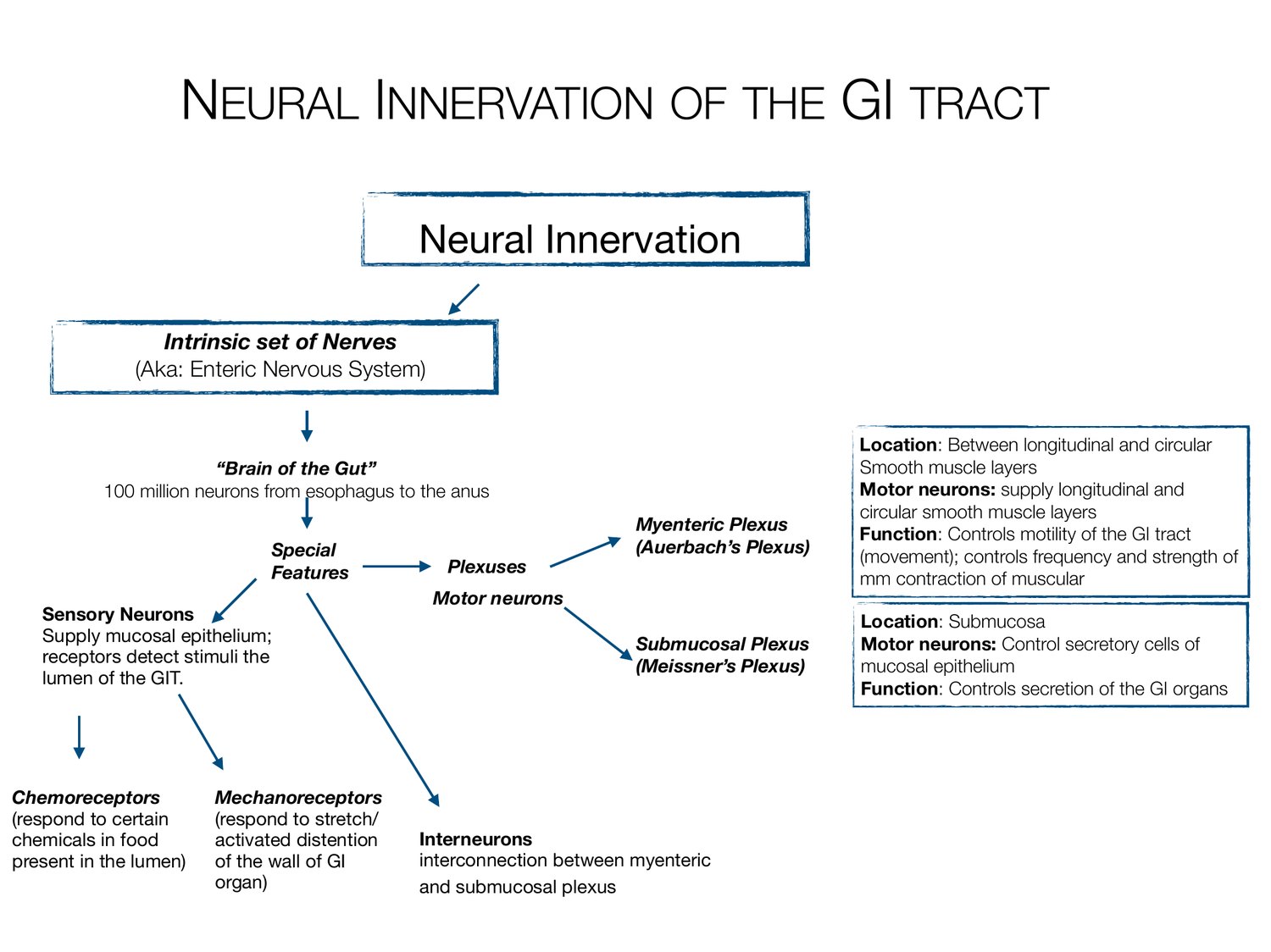
NEURAL INNERVATION OF THE GI TRACT Neural Innervation Intrinsic set of Nerves (Aka: Enteric Nervous System) Location: Between longitudinal and circular “Brain of the Gut” Smooth muscle layers 100 million neurons from esophagus to the anus Motor neurons: supply longitudinal and circular smooth muscle layers Myenteric Plexus Function: Controls motility of the GI tract Special (Auerbach’s Plexus) (movement); controls frequency and strength of Features Plexuses mm contraction of muscular Motor neurons Sensory Neurons Location: Submucosa Supply mucosal epithelium; Submucosal Plexus Motor neurons: Control secretory cells of receptors detect stimuli the (Meissner’s Plexus) mucosal epithelium lumen of the GIT. Function: Controls secretion of the GI organs Chemoreceptors Mechanoreceptors (respond to certain (respond to stretch/ chemicals in food activated distention Interneurons present in the lumen) of the wall of GI interconnection between myenteric organ) and submucosal plexus
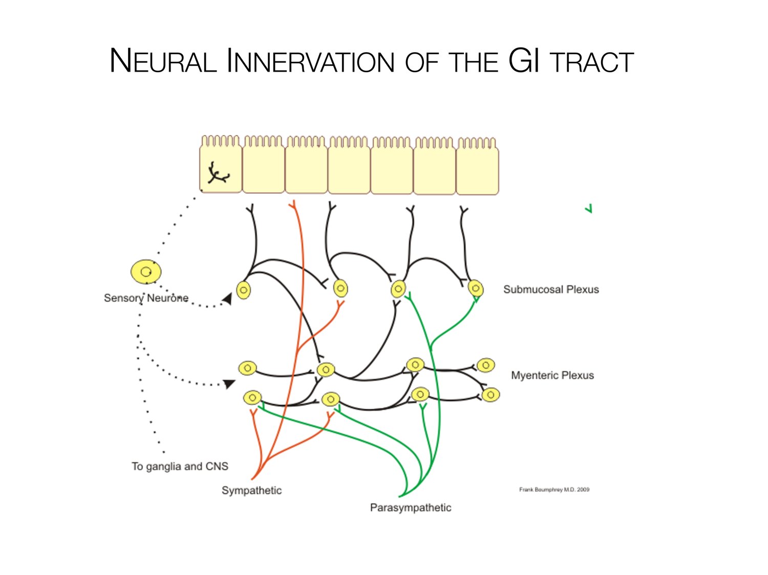
NEURAL INNERVATION OF THE GI TRACT
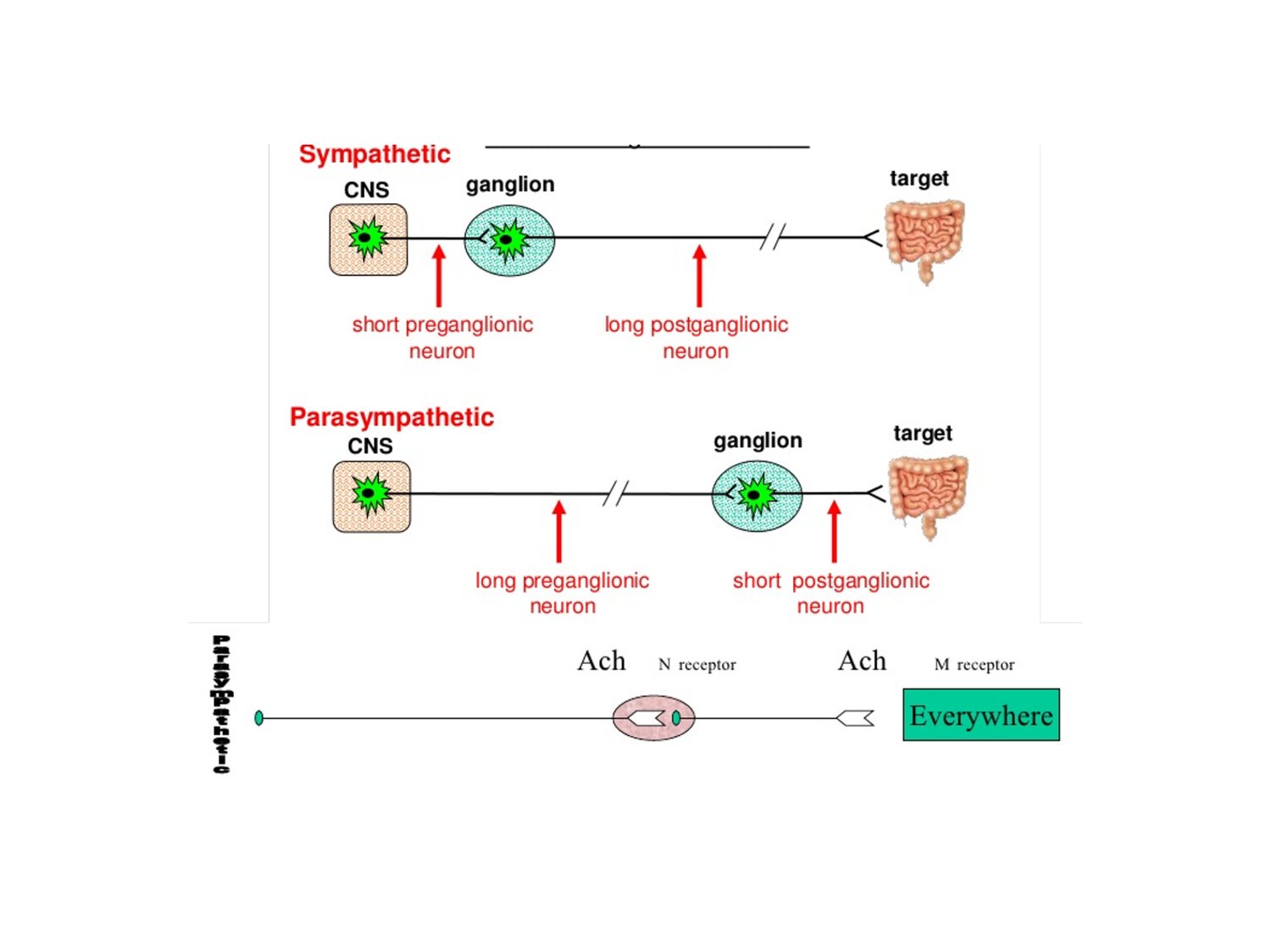
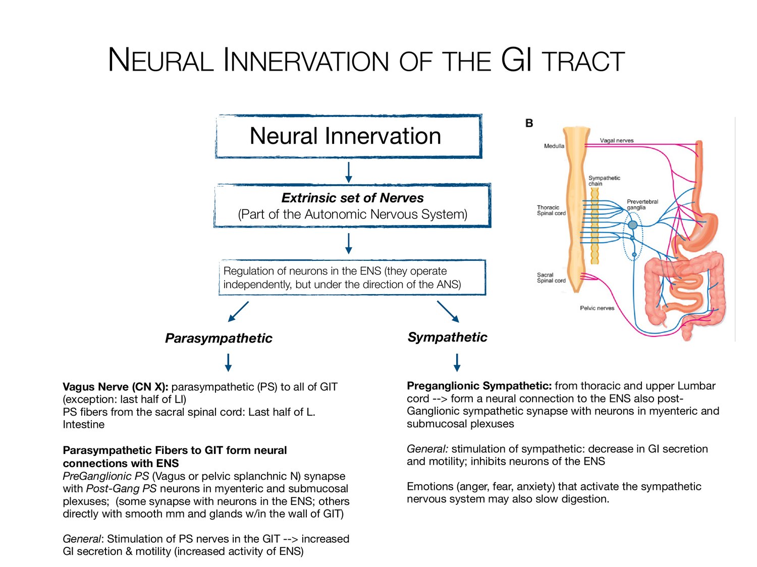
NEURAL INNERVATION OF THE GI TRACT Neural Innervation Extrinsic set of Nerves (Part of the Autonomic Nervous System) Regulation of neurons in the ENS (they operate independently, but under the direction of the ANS) Parasympathetic Sympathetic Vagus Nerve (CN X): parasympathetic (PS) to all of GIT Preganglionic Sympathetic: from thoracic and upper Lumbar (exception: last half of LI) cord --> form a neural connection to the ENS also post- PS bers from the sacral spinal cord: Last half of L. Ganglionic sympathetic synapse with neurons in myenteric and Intestine submucosal plexuses Parasympathetic Fibers to GIT form neural General: stimulation of sympathetic: decrease in GI secretion connections with ENS and motility; inhibits neurons of the ENS PreGanglionic PS (Vagus or pelvic splanchnic N) synapse with Post-Gang PS neurons in myenteric and submucosal Emotions (anger, fear, anxiety) that activate the sympathetic plexuses; (some synapse with neurons in the ENS; others nervous system may also slow digestion. directly with smooth mm and glands w/in the wall of GIT) General: Stimulation of PS nerves in the GIT -
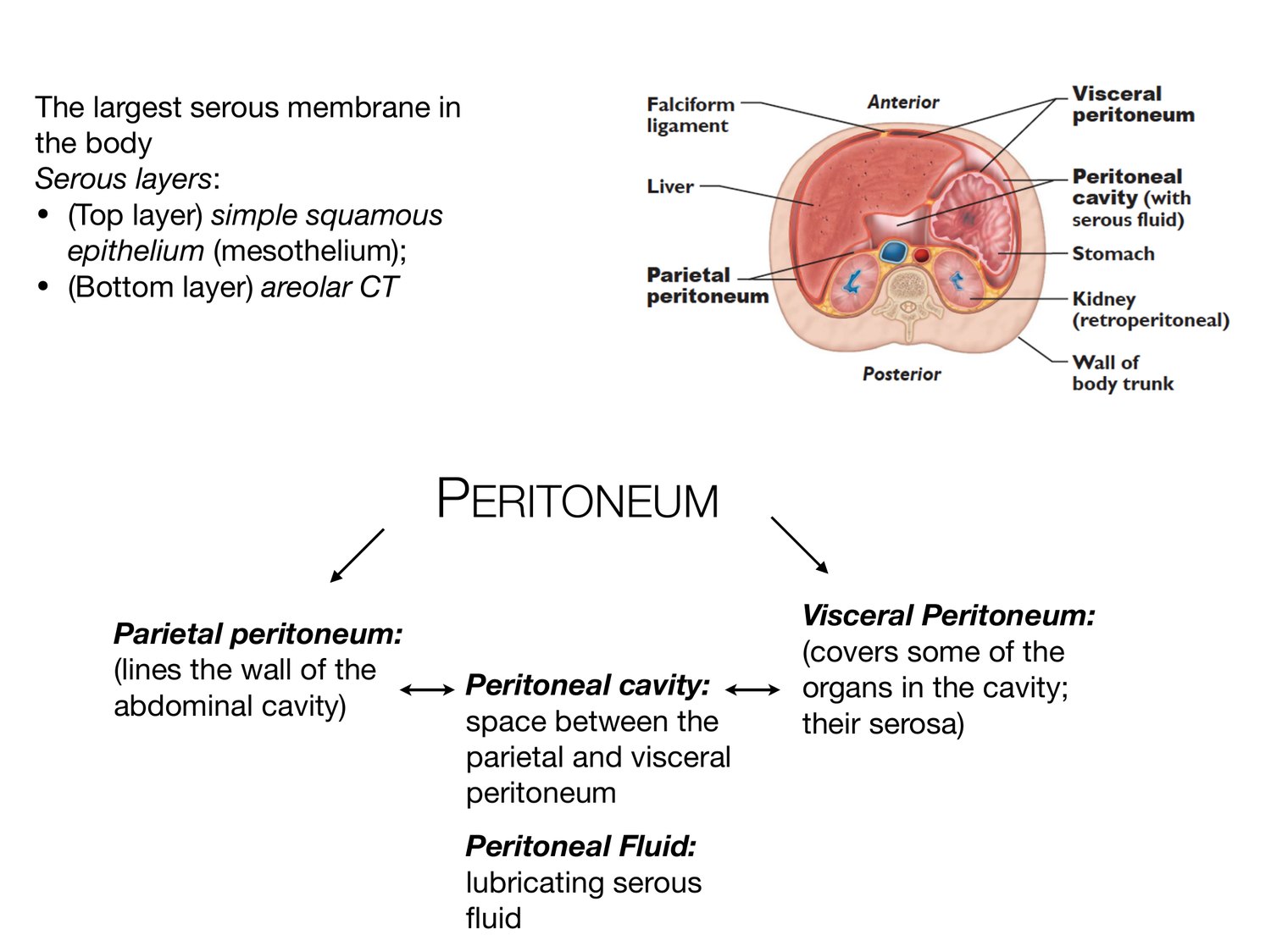
The largest serous membrane in the body Serous layers: • (Top layer) simple squamous epithelium (mesothelium); • (Bottom layer) areolar CT PERITONEUM Visceral Peritoneum: Parietal peritoneum: (covers some of the (lines the wall of the Peritoneal cavity: organs in the cavity; abdominal cavity) space between the their serosa) parietal and visceral peritoneum Peritoneal Fluid: lubricating serous uid fl
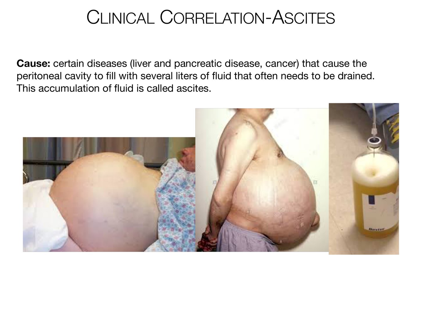
CLINICAL CORRELATION-ASCITES Cause: certain diseases (liver and pancreatic disease, cancer) that cause the peritoneal cavity to ll with several liters of uid that often needs to be drained. This accumulation of uid is called ascites. fi fl fl
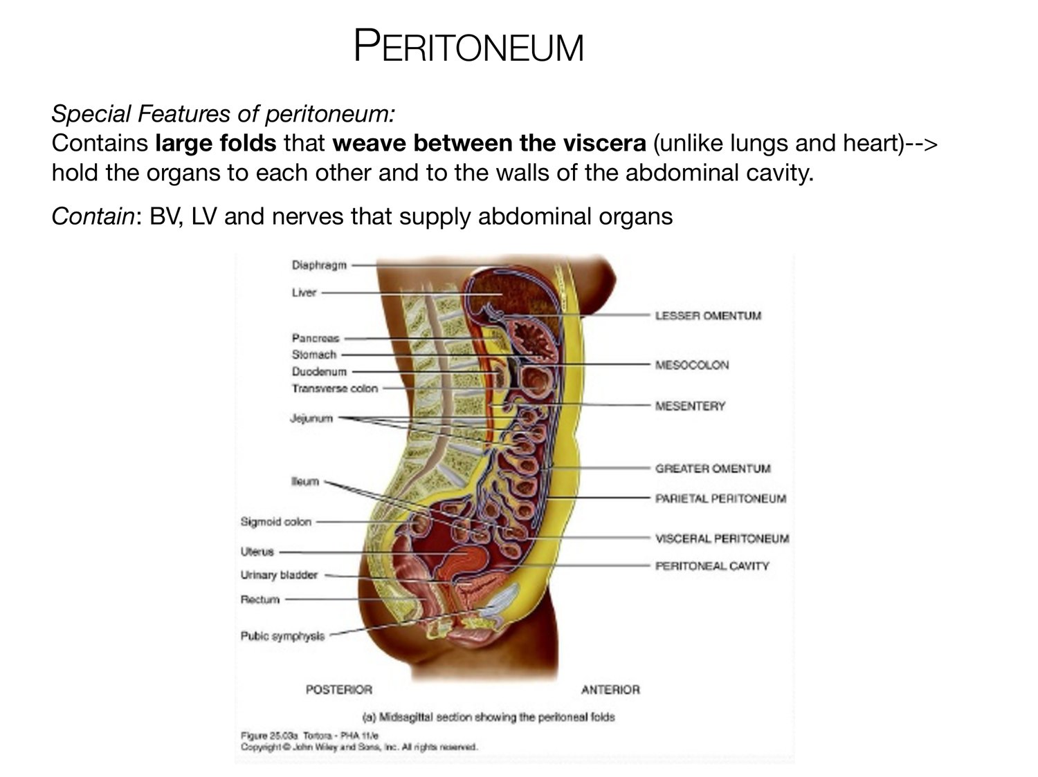
PERITONEUM Special Features of peritoneum: Contains large folds that weave between the viscera (unlike lungs and heart)--> hold the organs to each other and to the walls of the abdominal cavity. Contain: BV, LV and nerves that supply abdominal organs
FIVE PERITONEAL FOLDS-FALCIFORM LIGAMENT Falciform Ligament Location: Attaches the liver to the anterior abdominal wall and diaphragm. (The liver is the only organ that is attached to the anterior abdominal wall).
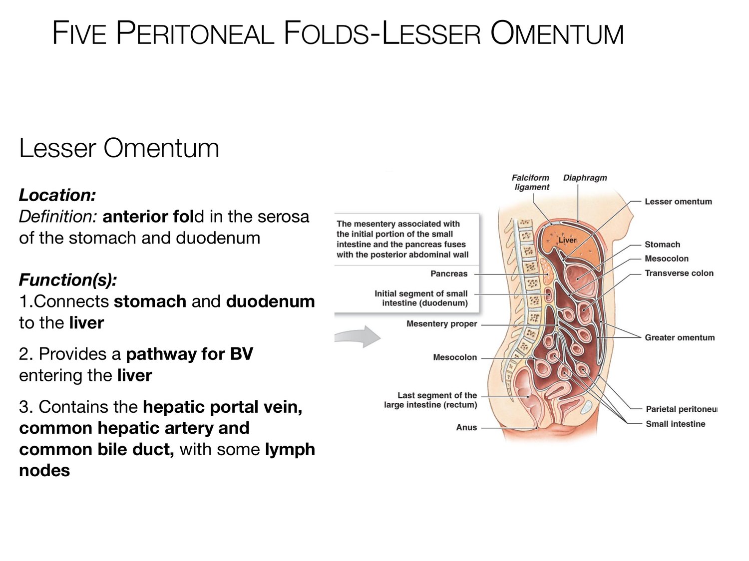
FIVE PERITONEAL FOLDS-LESSER OMENTUM Lesser Omentum Location: De nition: anterior fold in the serosa of the stomach and duodenum Function(s): 1.Connects stomach and duodenum to the liver 2. Provides a pathway for BV entering the liver 3. Contains the hepatic portal vein, common hepatic artery and common bile duct, with some lymph nodes fi
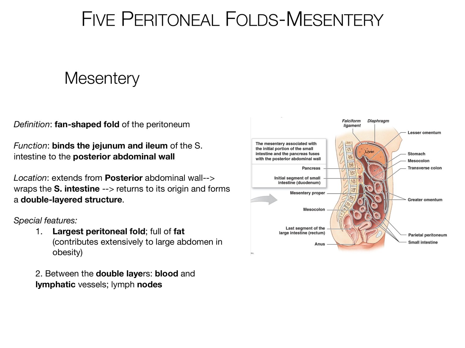
FIVE PERITONEAL FOLDS-MESENTERY Mesentery De nition: fan-shaped fold of the peritoneum Function: binds the jejunum and ileum of the S. intestine to the posterior abdominal wall Location: extends from Posterior abdominal wall--> wraps the S. intestine --> returns to its origin and forms a double-layered structure. Special features: 1. Largest peritoneal fold; full of fat (contributes extensively to large abdomen in obesity) 2. Between the double layers: blood and lymphatic vessels; lymph nodes fi
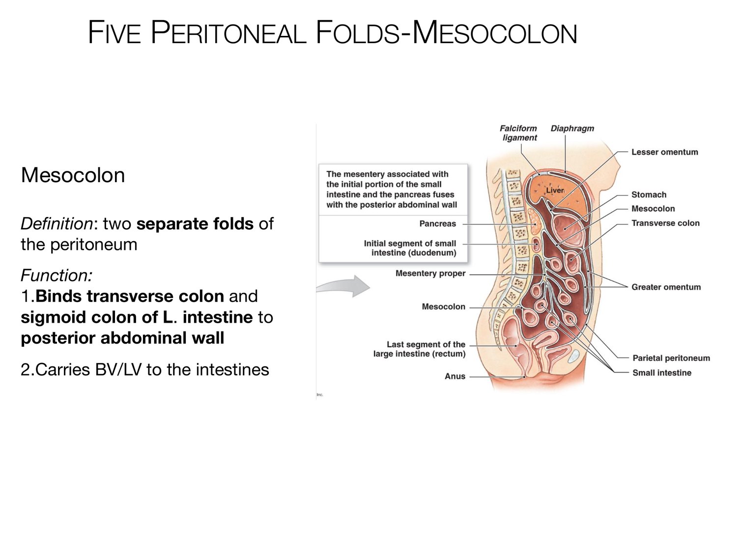
FIVE PERITONEAL FOLDS-MESOCOLON Mesocolon De nition: two separate folds of the peritoneum Function: 1.Binds transverse colon and sigmoid colon of L. intestine to posterior abdominal wall 2.Carries BV/LV to the intestines fi
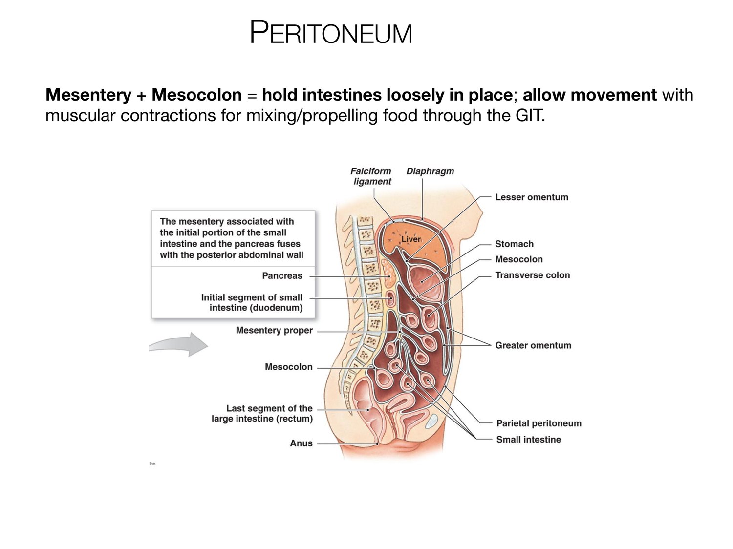
PERITONEUM Mesentery + Mesocolon = hold intestines loosely in place; allow movement with muscular contractions for mixing/propelling food through the GIT.
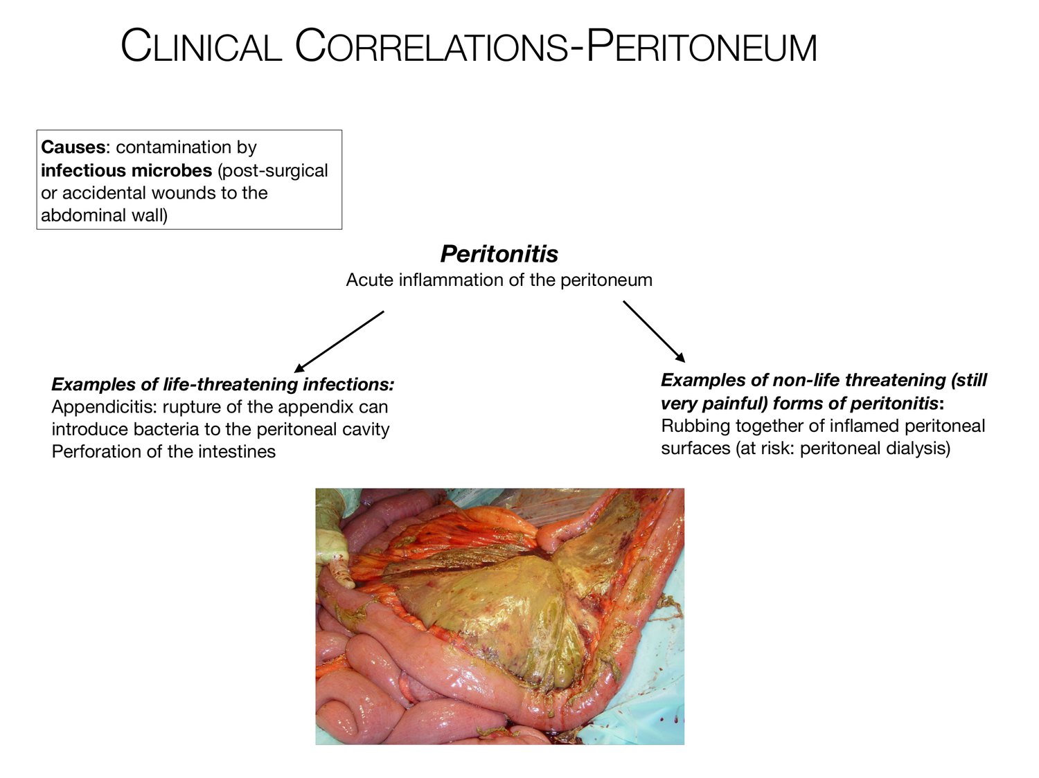
CLINICAL CORRELATIONS-PERITONEUM Causes: contamination by infectious microbes (post-surgical or accidental wounds to the abdominal wall) Peritonitis Acute in ammation of the peritoneum Examples of life-threatening infections: Examples of non-life threatening (still Appendicitis: rupture of the appendix can very painful) forms of peritonitis: introduce bacteria to the peritoneal cavity Rubbing together of in amed peritoneal Perforation of the intestines surfaces (at risk: peritoneal dialysis) fl fl
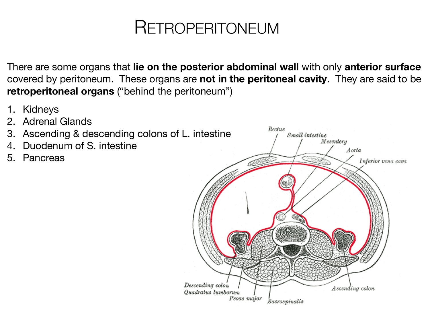
RETROPERITONEUM There are some organs that lie on the posterior abdominal wall with only anterior surface covered by peritoneum. These organs are not in the peritoneal cavity. They are said to be retroperitoneal organs (“behind the peritoneum”) 1. Kidneys 2. Adrenal Glands 3. Ascending & descending colons of L. intestine 4. Duodenum of S. intestine 5. Pancreas
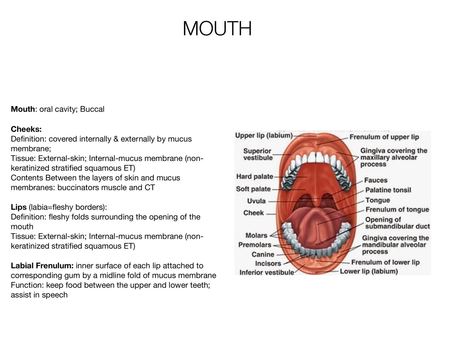
MOUTH Mouth: oral cavity; Buccal Cheeks: De nition: covered internally & externally by mucus membrane; Tissue: External-skin; Internal-mucus membrane (non- keratinized strati ed squamous ET) Contents Between the layers of skin and mucus membranes: buccinators muscle and CT Lips (labia= eshy borders): De nition: eshy folds surrounding the opening of the mouth Tissue: External-skin; Internal-mucus membrane (non- keratinized strati ed squamous ET) Labial Frenulum: inner surface of each lip attached to corresponding gum by a midline fold of mucus membrane Function: keep food between the upper and lower teeth; assist in speech fi fi fl fl fi fi
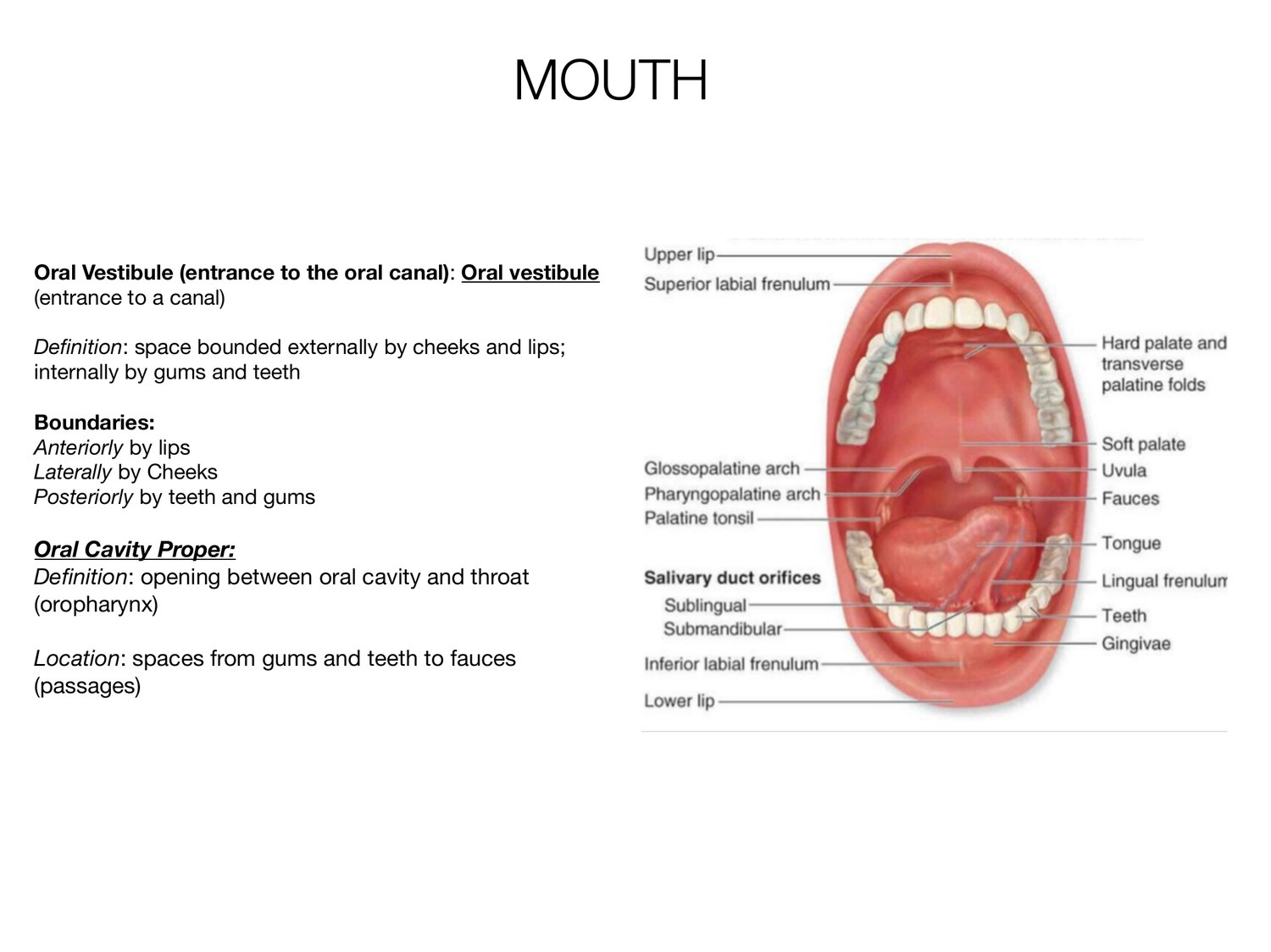
MOUTH Oral Vestibule (entrance to the oral canal): Oral vestibule (entrance to a canal) De nition: space bounded externally by cheeks and lips; internally by gums and teeth Boundaries: Anteriorly by lips Laterally by Cheeks Posteriorly by teeth and gums Oral Cavity Proper: De nition: opening between oral cavity and throat (oropharynx) Location: spaces from gums and teeth to fauces (passages) fi fi
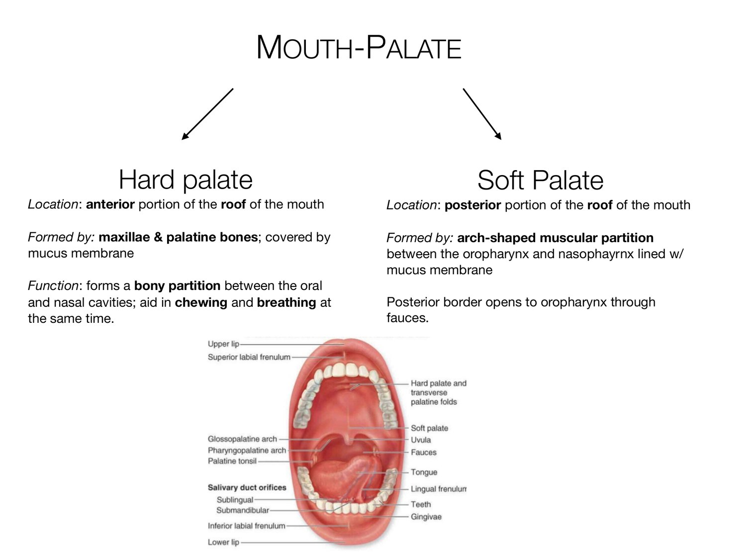
MOUTH-PALATE Hard palate Soft Palate Location: anterior portion of the roof of the mouth Location: posterior portion of the roof of the mouth Formed by: maxillae & palatine bones; covered by Formed by: arch-shaped muscular partition mucus membrane between the oropharynx and nasophayrnx lined w/ mucus membrane Function: forms a bony partition between the oral and nasal cavities; aid in chewing and breathing at Posterior border opens to oropharynx through the same time. fauces.
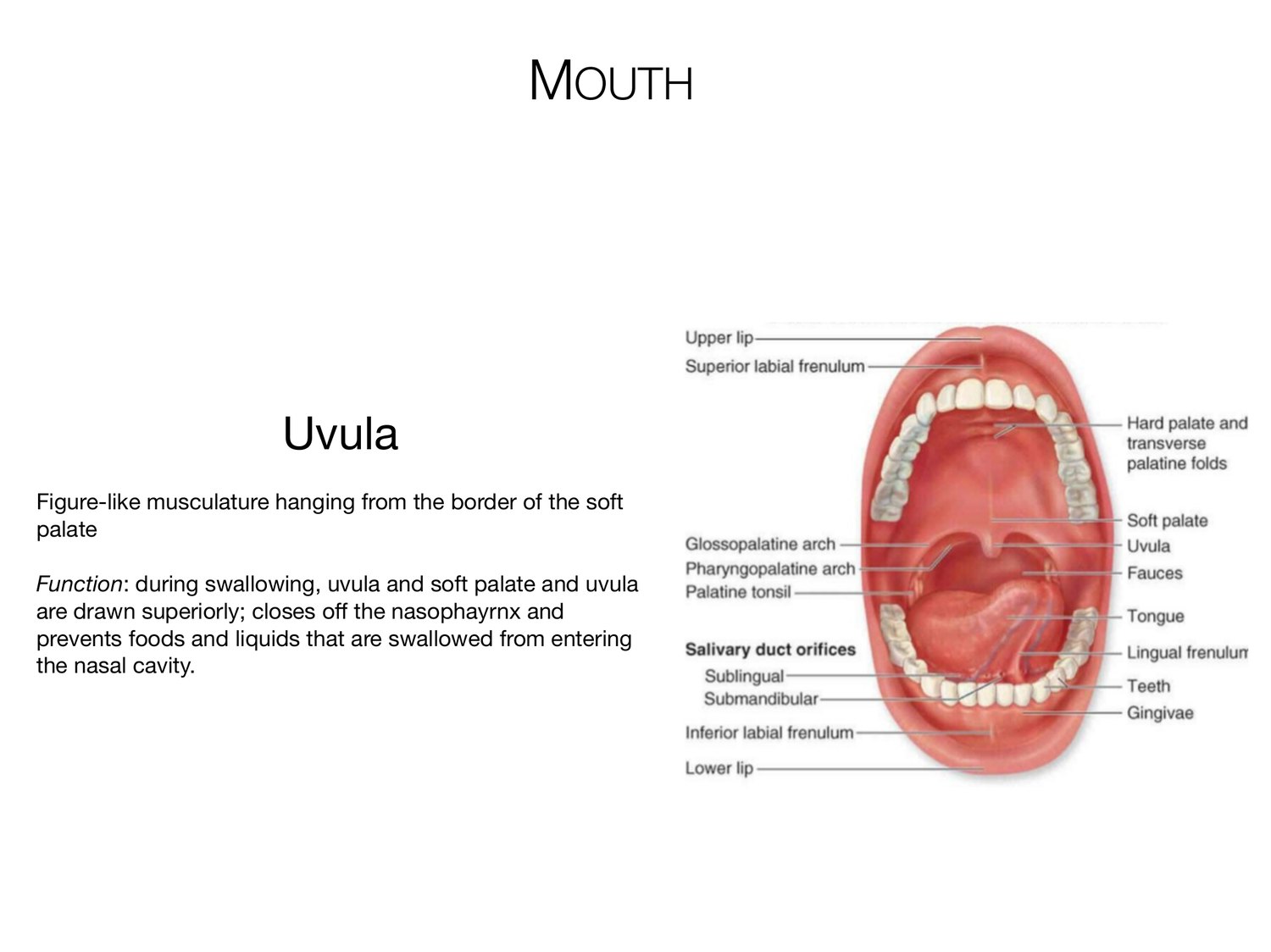
MOUTH Uvula Figure-like musculature hanging from the border of the soft palate Function: during swallowing, uvula and soft palate and uvula are drawn superiorly; closes o the nasophayrnx and prevents foods and liquids that are swallowed from entering the nasal cavity. ff
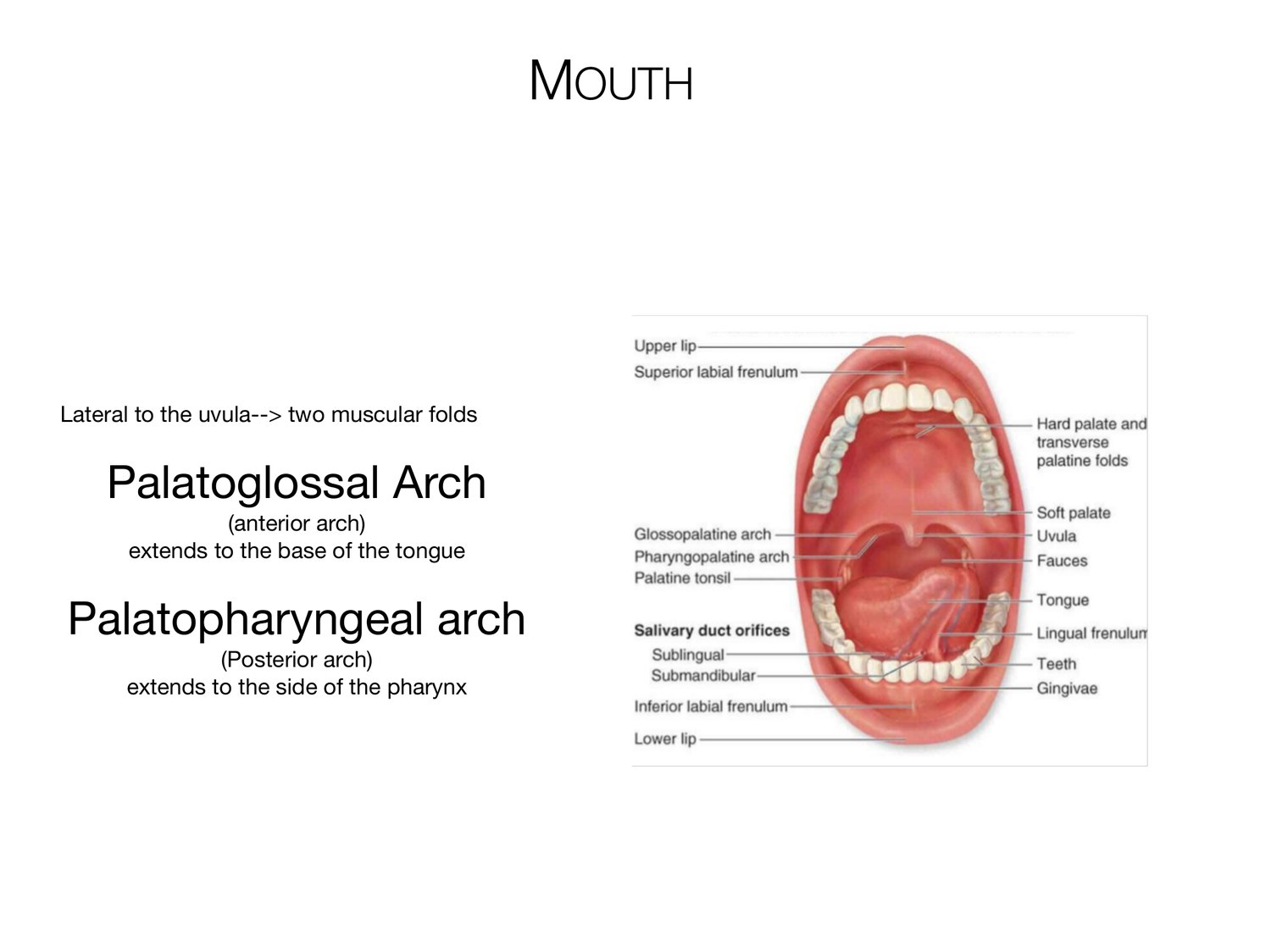
MOUTH Lateral to the uvula--> two muscular folds Palatoglossal Arch (anterior arch) extends to the base of the tongue Palatopharyngeal arch (Posterior arch) extends to the side of the pharynx
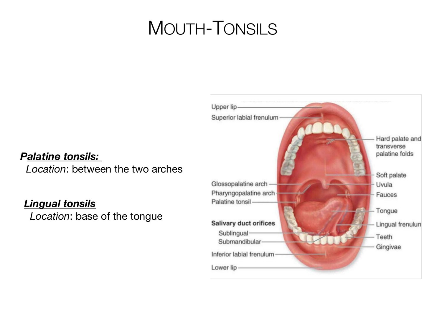
MOUTH-TONSILS Palatine tonsils: Location: between the two arches Lingual tonsils Location: base of the tongue
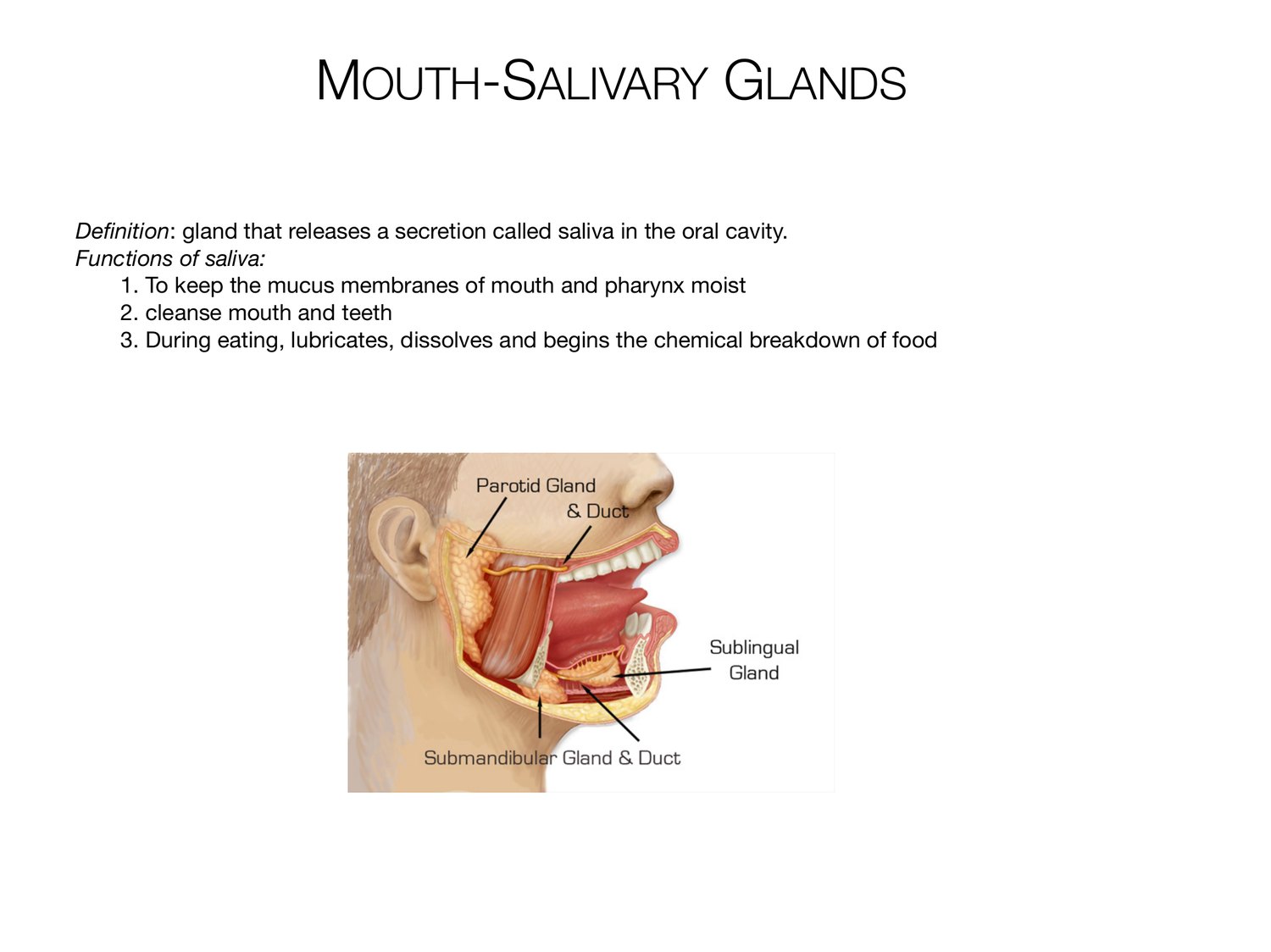
MOUTH-SALIVARY GLANDS Definition: gland that releases a secretion called saliva in the oral cavity. Functions of saliva: 1. To keep the mucus membranes of mouth and pharynx moist 2. cleanse mouth and teeth 3. During eating, lubricates, dissolves and begins the chemical breakdown of food
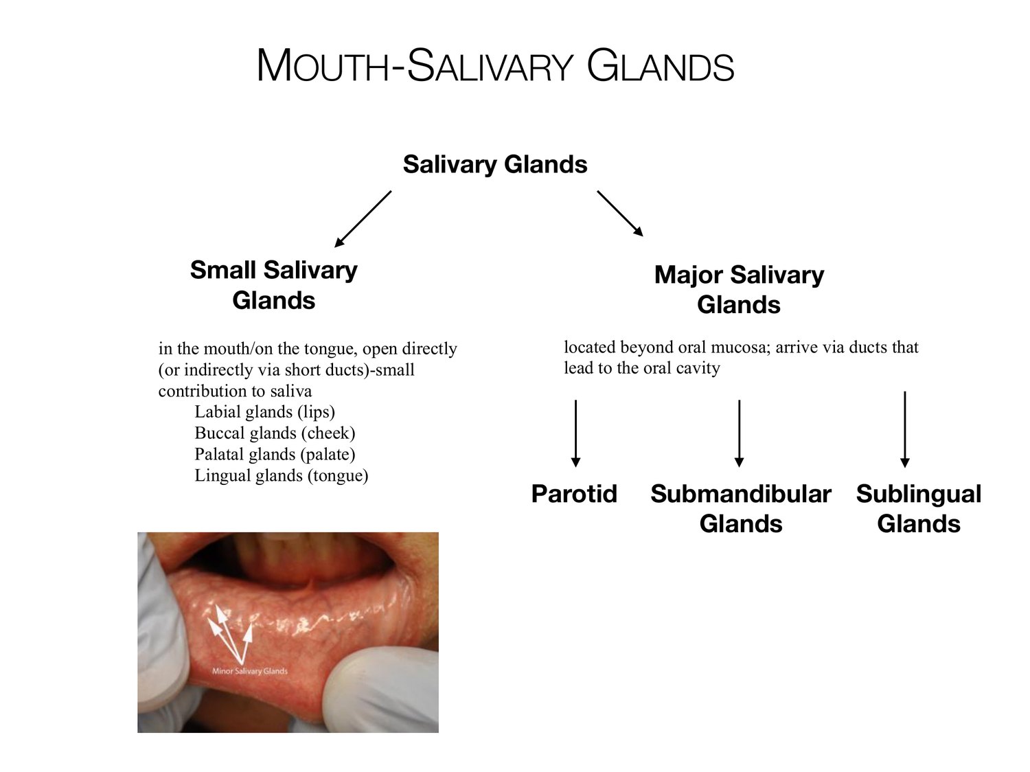
MOUTH-SALIVARY GLANDS Salivary Glands Small Salivary Major Salivary Glands Glands in the mouth/on the tongue, open directly located beyond oral mucosa; arrive via ducts that (or indirectly via short ducts)-small lead to the oral cavity contribution to saliva Labial glands (lips) Buccal glands (cheek) Palatal glands (palate) Lingual glands (tongue) Parotid Submandibular Sublingual Glands Glands
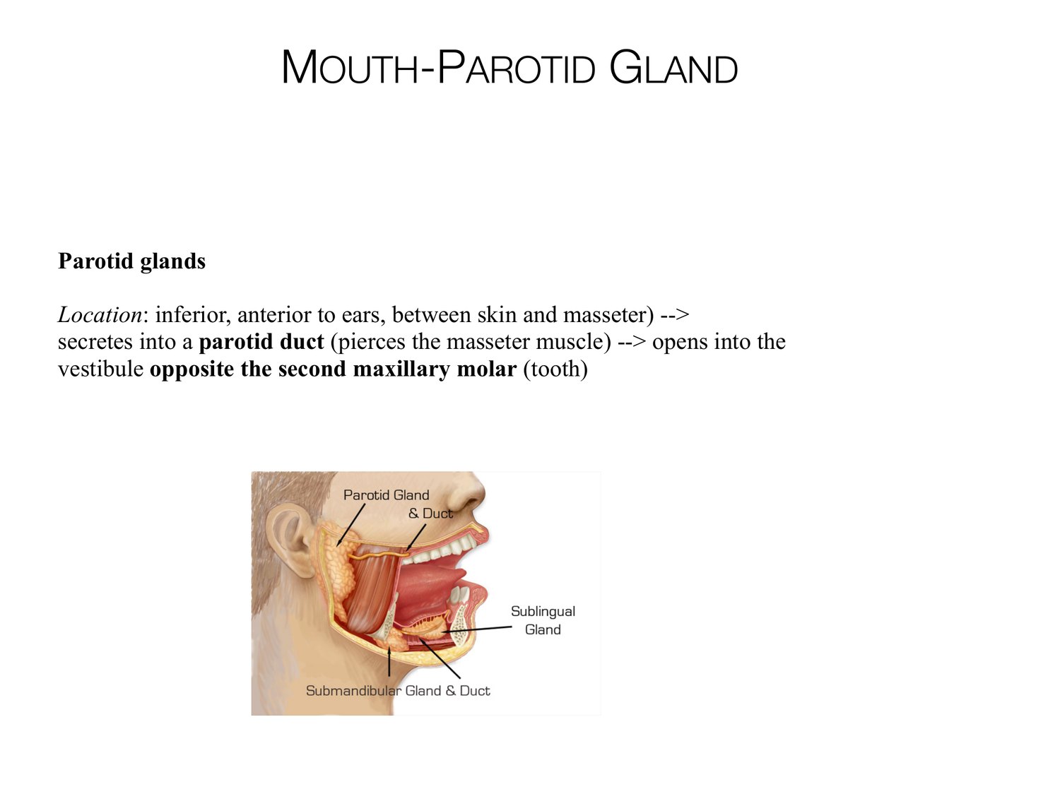
MOUTH-PAROTID GLAND Parotid glands Location: inferior, anterior to ears, between skin and masseter) --> secretes into a parotid duct (pierces the masseter muscle) --> opens into the vestibule opposite the second maxillary molar (tooth)
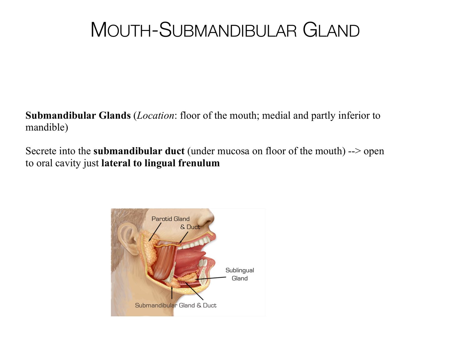
MOUTH-SUBMANDIBULAR GLAND Submandibular Glands (Location: floor of the mouth; medial and partly inferior to mandible) Secrete into the submandibular duct (under mucosa on floor of the mouth) --> open to oral cavity just lateral to lingual frenulum
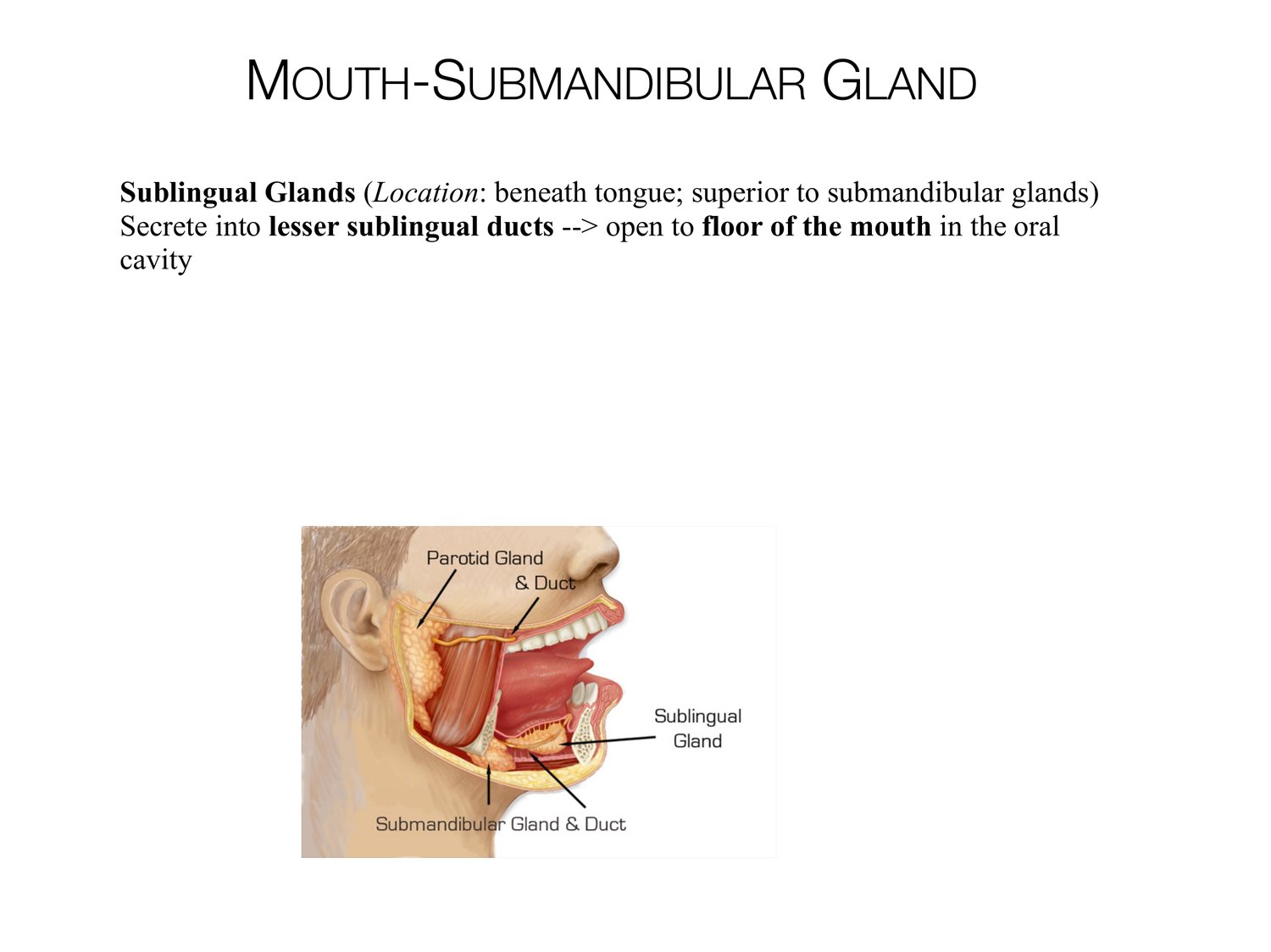
MOUTH-SUBMANDIBULAR GLAND Sublingual Glands (Location: beneath tongue; superior to submandibular glands) Secrete into lesser sublingual ducts --> open to floor of the mouth in the oral cavity
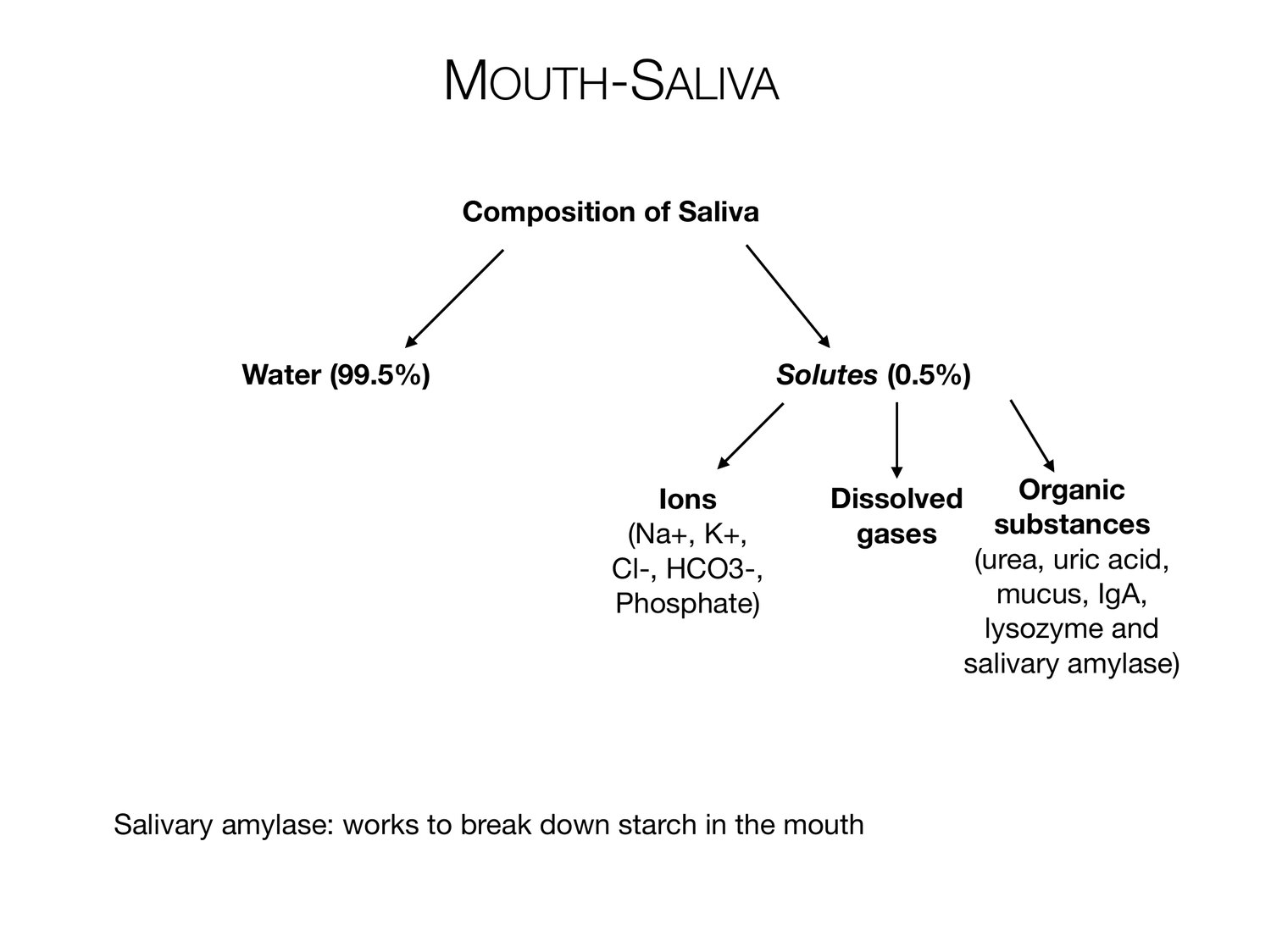
MOUTH-SALIVA Composition of Saliva Water (99.5%) Solutes (0.5%) Ions Dissolved Organic (Na+, K+, gases substances Cl-, HCO3-, (urea, uric acid, Phosphate) mucus, IgA, lysozyme and salivary amylase) Salivary amylase: works to break down starch in the mouth
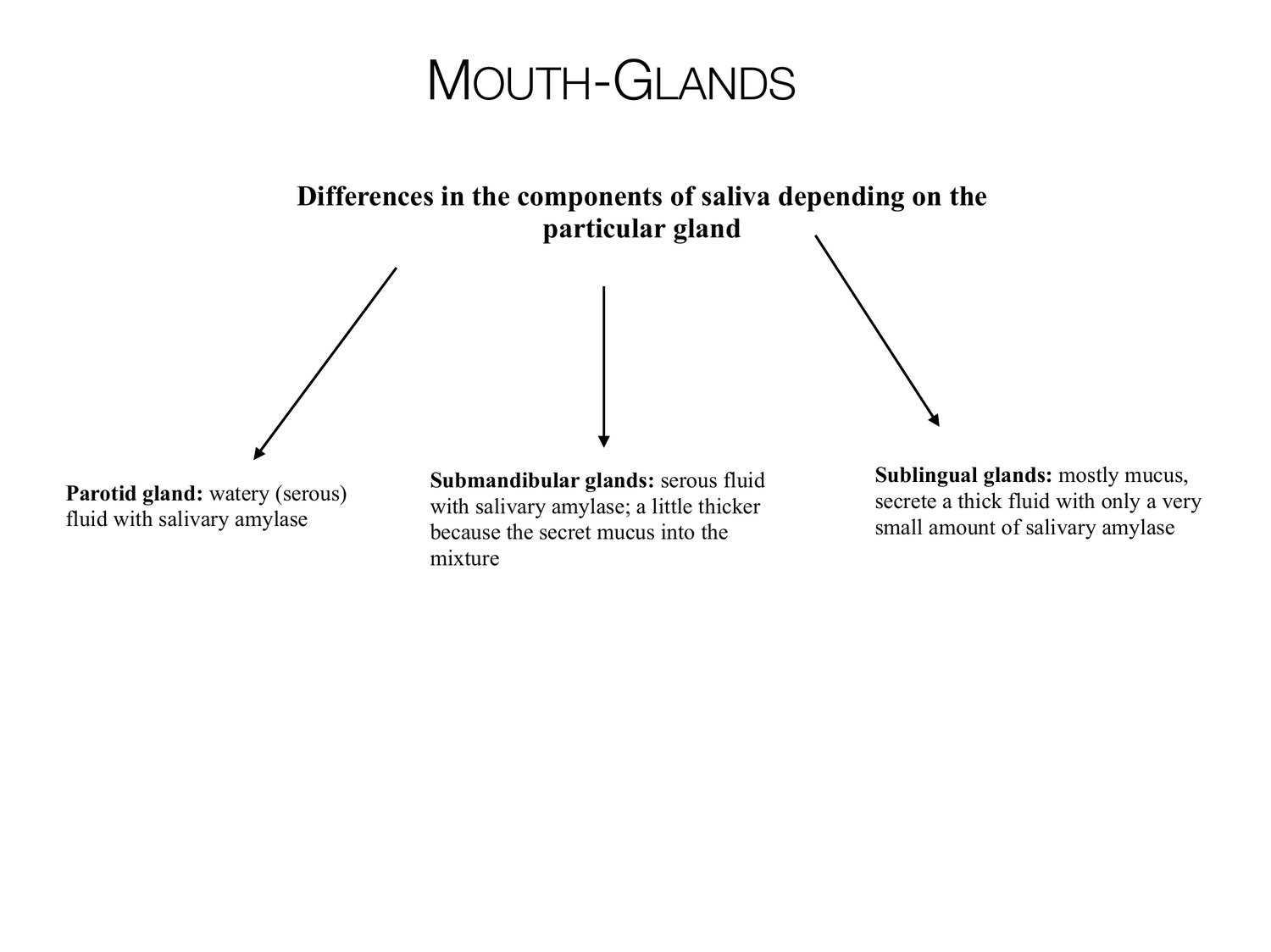
MOUTH-GLANDS Differences in the components of saliva depending on the particular gland Submandibular glands: serous fluid Sublingual glands: mostly mucus, Parotid gland: watery (serous) secrete a thick fluid with only a very with salivary amylase; a little thicker fluid with salivary amylase small amount of salivary amylase because the secret mucus into the mixture
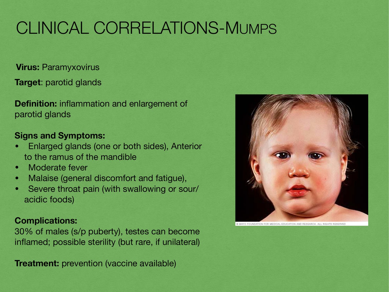
CLINICAL CORRELATIONS-MUMPS Virus: Paramyxovirus Target: parotid glands Definition: inflammation and enlargement of parotid glands Signs and Symptoms: • Enlarged glands (one or both sides), Anterior to the ramus of the mandible • Moderate fever • Malaise (general discomfort and fatigue), • Severe throat pain (with swallowing or sour/ acidic foods) Complications: 30% of males (s/p puberty), testes can become inflamed; possible sterility (but rare, if unilateral) Treatment: prevention (vaccine available)
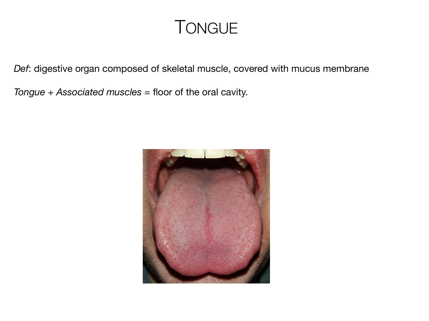
TONGUE Def: digestive organ composed of skeletal muscle, covered with mucus membrane Tongue + Associated muscles = floor of the oral cavity.
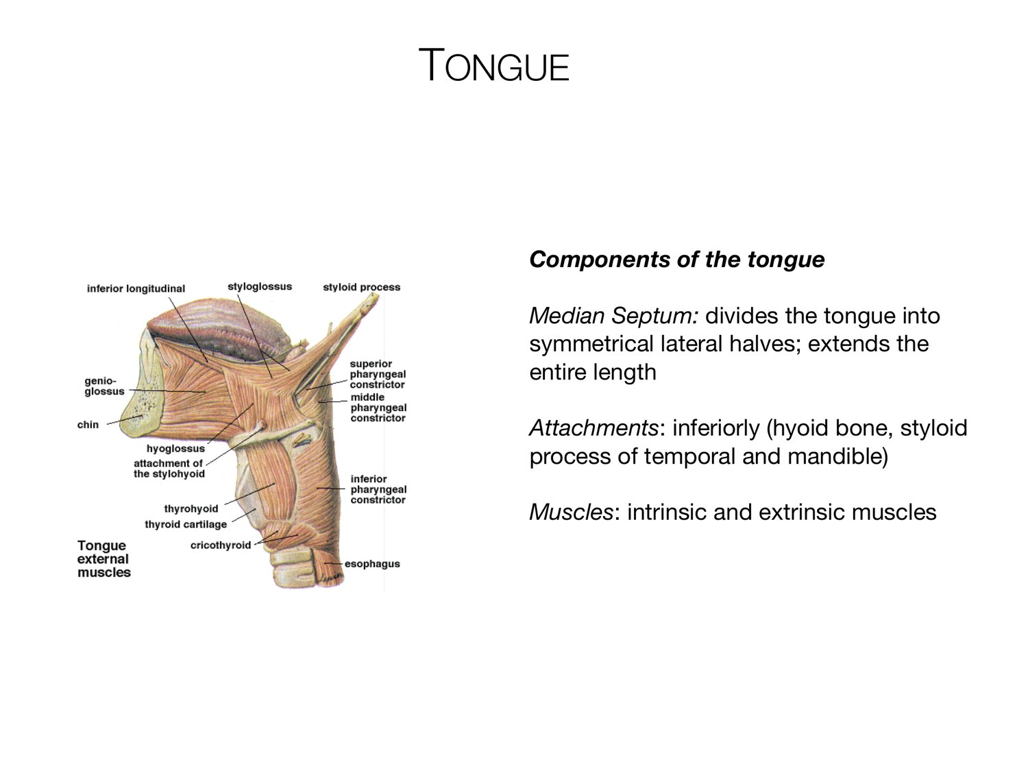
TONGUE Components of the tongue Median Septum: divides the tongue into symmetrical lateral halves; extends the entire length Attachments: inferiorly (hyoid bone, styloid process of temporal and mandible) Muscles: intrinsic and extrinsic muscles
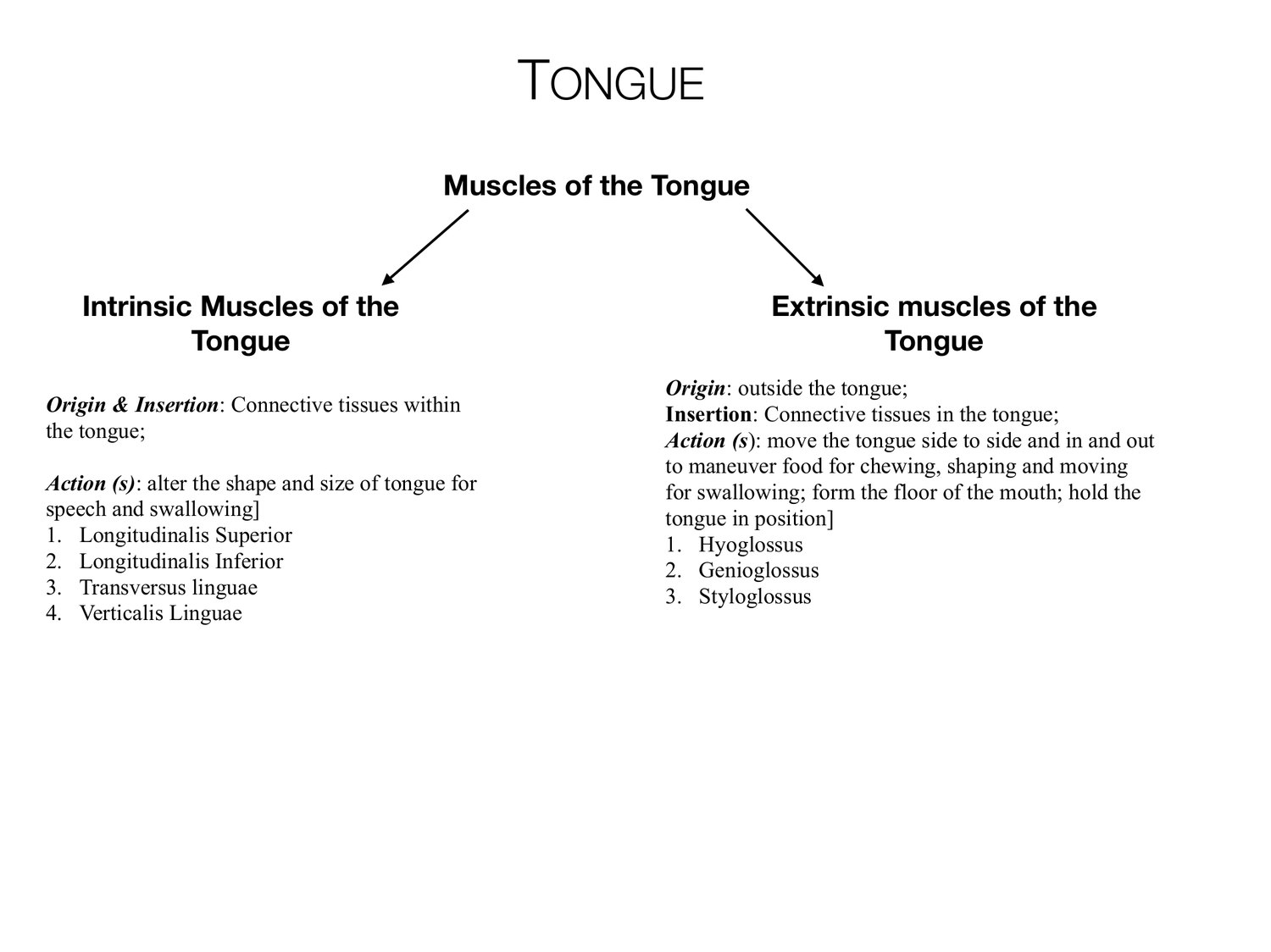
TONGUE Muscles of the Tongue Intrinsic Muscles of the Extrinsic muscles of the Tongue Tongue Origin: outside the tongue; Origin & Insertion: Connective tissues within Insertion: Connective tissues in the tongue; the tongue; Action (s): move the tongue side to side and in and out to maneuver food for chewing, shaping and moving Action (s): alter the shape and size of tongue for for swallowing; form the floor of the mouth; hold the speech and swallowing] tongue in position] 1. Longitudinalis Superior 1. Hyoglossus 2. Longitudinalis Inferior 2. Genioglossus 3. Transversus linguae 3. Styloglossus 4. Verticalis Linguae
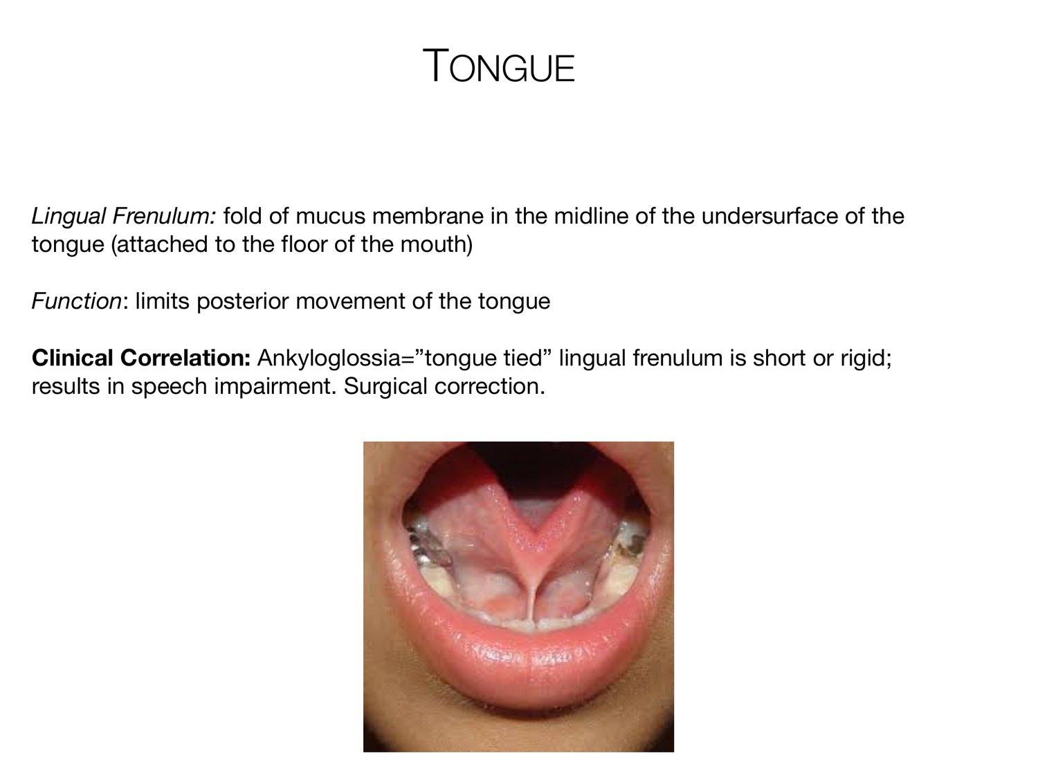
TONGUE Lingual Frenulum: fold of mucus membrane in the midline of the undersurface of the tongue (attached to the floor of the mouth) Function: limits posterior movement of the tongue Clinical Correlation: Ankyloglossia=”tongue tied” lingual frenulum is short or rigid; results in speech impairment. Surgical correction.
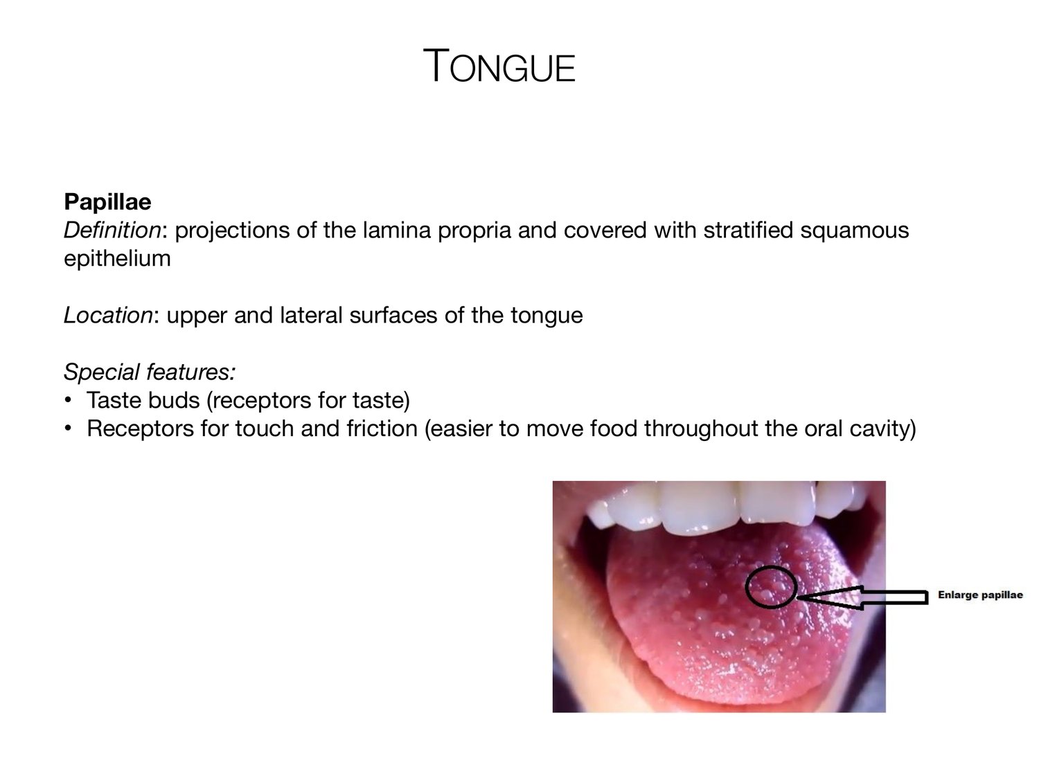
TONGUE Papillae Definition: projections of the lamina propria and covered with stratified squamous epithelium Location: upper and lateral surfaces of the tongue Special features: • Taste buds (receptors for taste) • Receptors for touch and friction (easier to move food throughout the oral cavity)
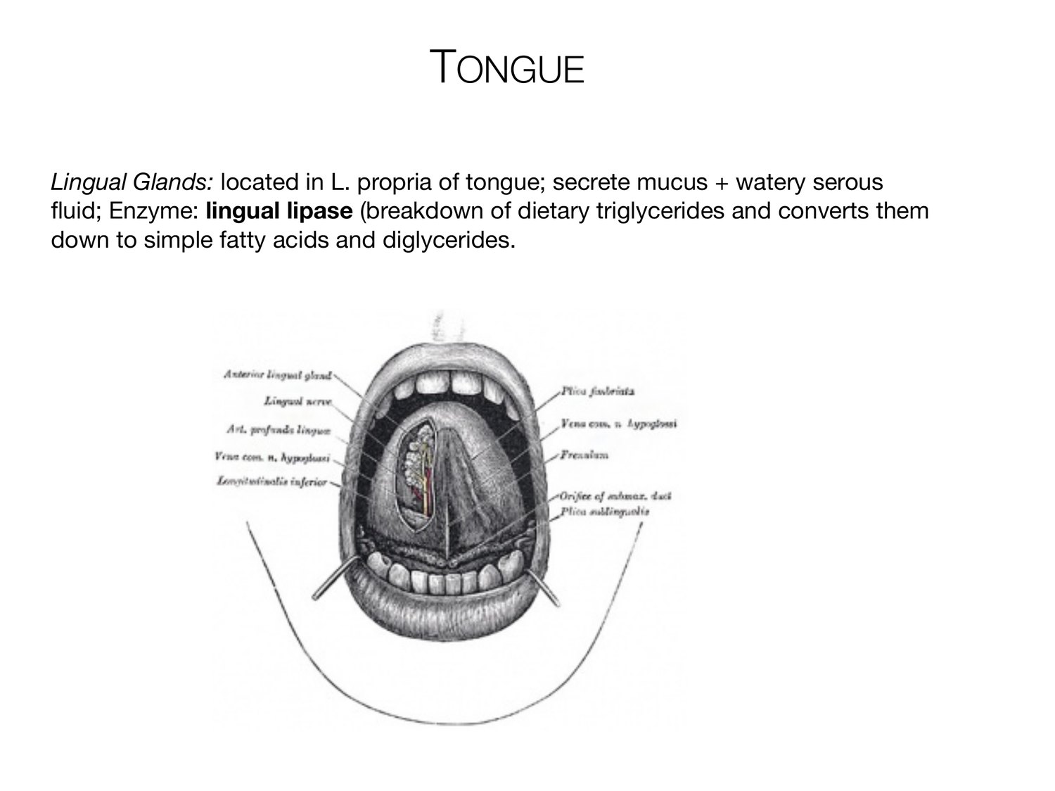
TONGUE Lingual Glands: located in L. propria of tongue; secrete mucus + watery serous fluid; Enzyme: lingual lipase (breakdown of dietary triglycerides and converts them down to simple fatty acids and diglycerides.
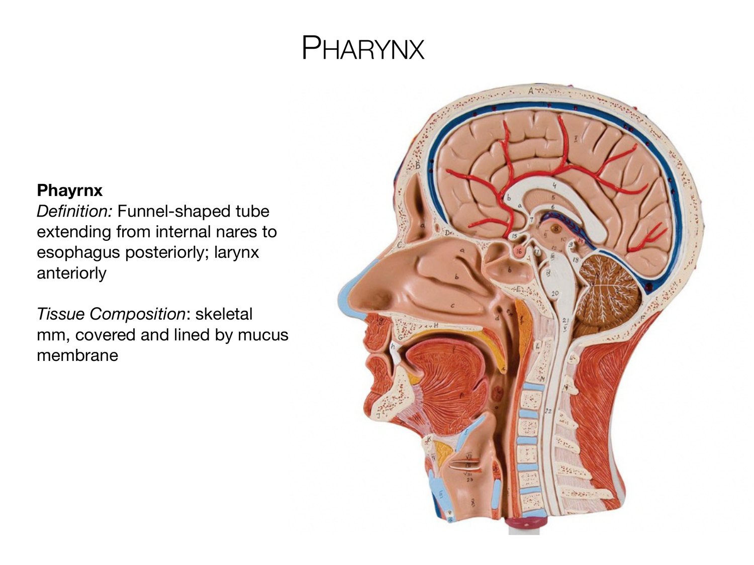
PHARYNX Phayrnx Definition: Funnel-shaped tube extending from internal nares to esophagus posteriorly; larynx anteriorly Tissue Composition: skeletal mm, covered and lined by mucus membrane
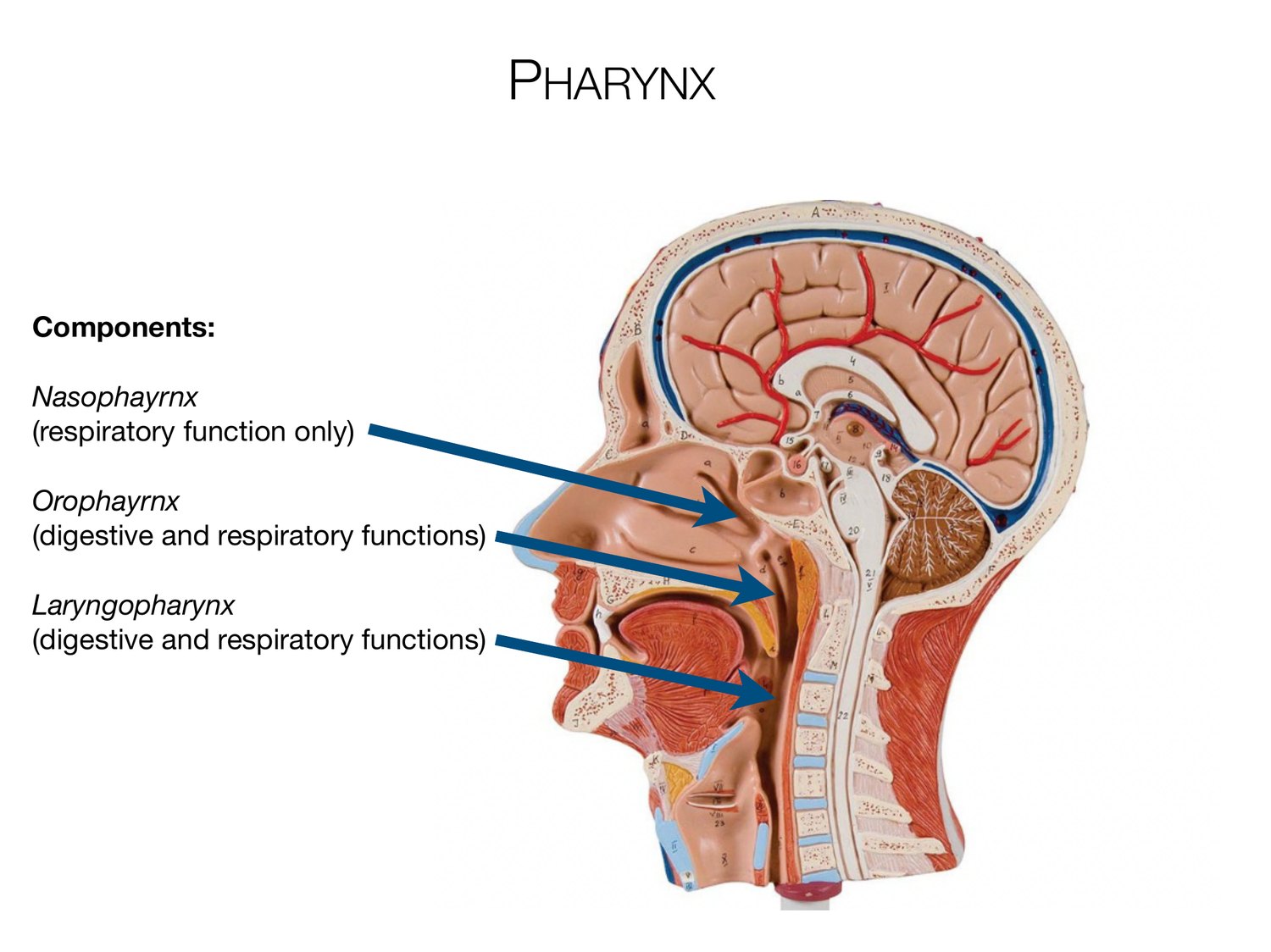
PHARYNX Components: Nasophayrnx (respiratory function only) Orophayrnx (digestive and respiratory functions) Laryngopharynx (digestive and respiratory functions)
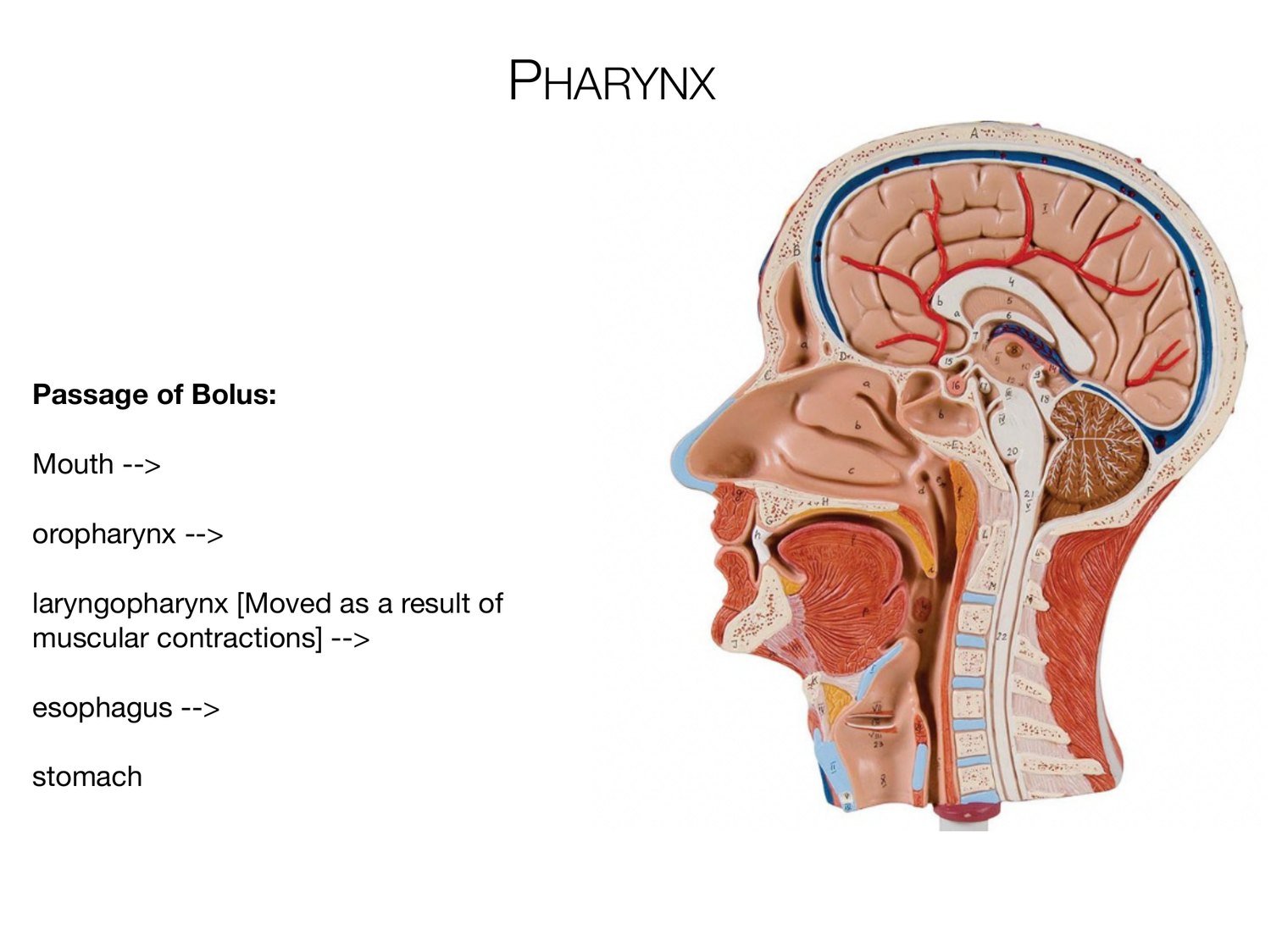
PHARYNX Passage of Bolus: Mouth --> oropharynx --> laryngopharynx [Moved as a result of muscular contractions] --> esophagus --> stomach
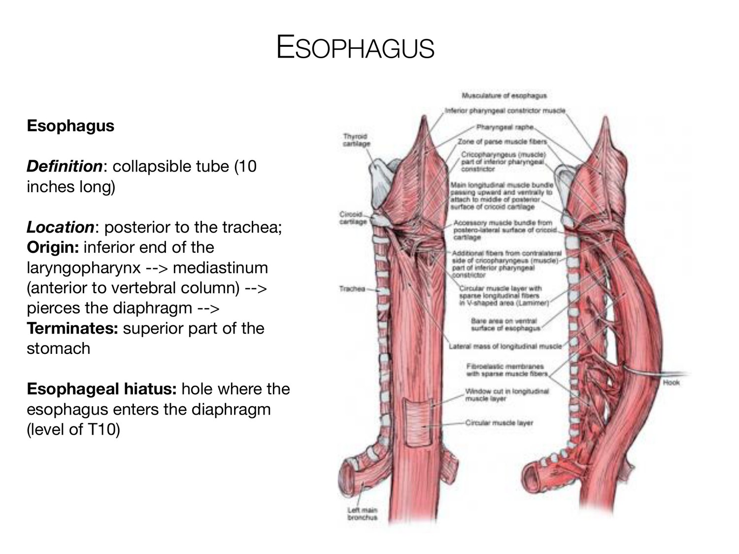
ESOPHAGUS Esophagus Definition: collapsible tube (10 inches long) Location: posterior to the trachea; Origin: inferior end of the laryngopharynx --> mediastinum (anterior to vertebral column) --> pierces the diaphragm --> Terminates: superior part of the stomach Esophageal hiatus: hole where the esophagus enters the diaphragm (level of T10)
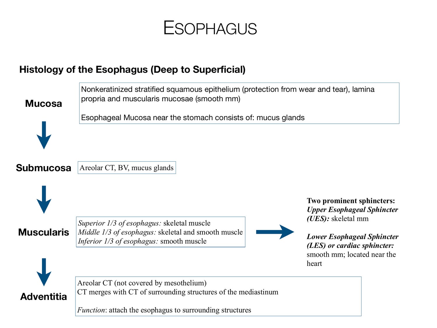
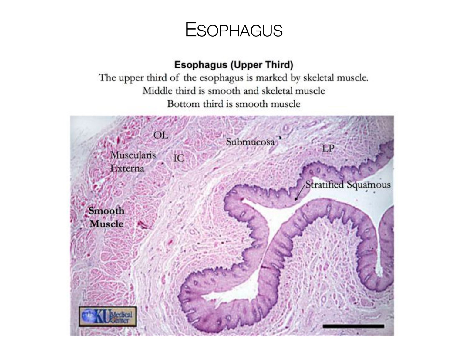
ESOPHAGUS Histology of the Esophagus (Deep to Superficial) Nonkeratinized stratified squamous epithelium (protection from wear and tear), lamina propria and muscularis mucosae (smooth mm) Mucosa Esophageal Mucosa near the stomach consists of: mucus glands Submucosa Areolar CT, BV, mucus glands Two prominent sphincters: Upper Esophageal Sphincter (UES): skeletal mm Superior 1/3 of esophagus: skeletal muscle Muscularis Middle 1/3 of esophagus: skeletal and smooth muscle Lower Esophageal Sphincter Inferior 1/3 of esophagus: smooth muscle (LES) or cardiac sphincter: smooth mm; located near the heart Areolar CT (not covered by mesothelium) CT merges with CT of surrounding structures of the mediastinum Adventitia Function: attach the esophagus to surrounding structures
ESOPHAGUS
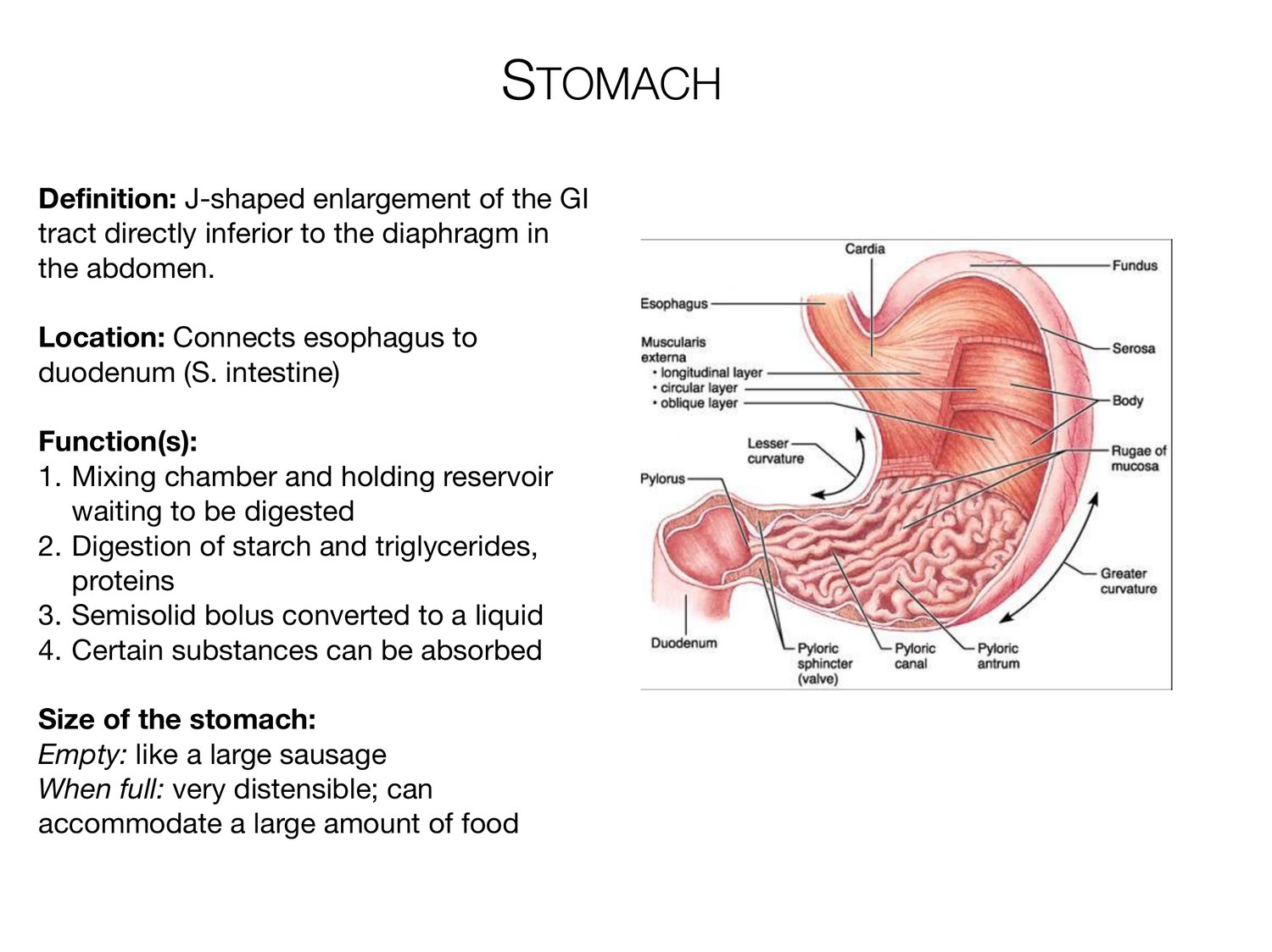
STOMACH Definition: J-shaped enlargement of the GI tract directly inferior to the diaphragm in the abdomen. Location: Connects esophagus to duodenum (S. intestine) Function(s): 1. Mixing chamber and holding reservoir waiting to be digested 2. Digestion of starch and triglycerides, proteins 3. Semisolid bolus converted to a liquid 4. Certain substances can be absorbed Size of the stomach: Empty: like a large sausage When full: very distensible; can accommodate a large amount of food
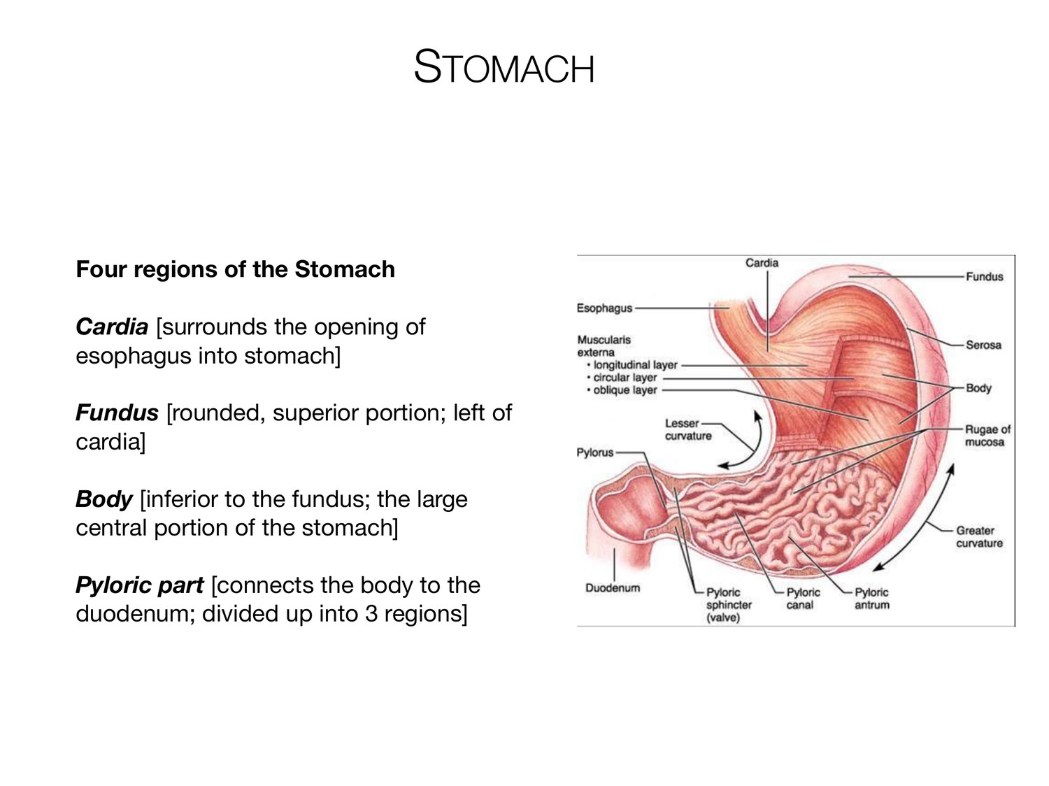
STOMACH Four regions of the Stomach Cardia [surrounds the opening of esophagus into stomach] Fundus [rounded, superior portion; left of cardia] Body [inferior to the fundus; the large central portion of the stomach] Pyloric part [connects the body to the duodenum; divided up into 3 regions]
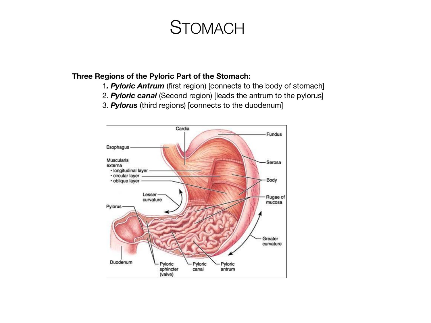
STOMACH Three Regions of the Pyloric Part of the Stomach: 1. Pyloric Antrum (first region) [connects to the body of stomach] 2. Pyloric canal (Second region) [leads the antrum to the pylorus] 3. Pylorus (third regions) [connects to the duodenum]
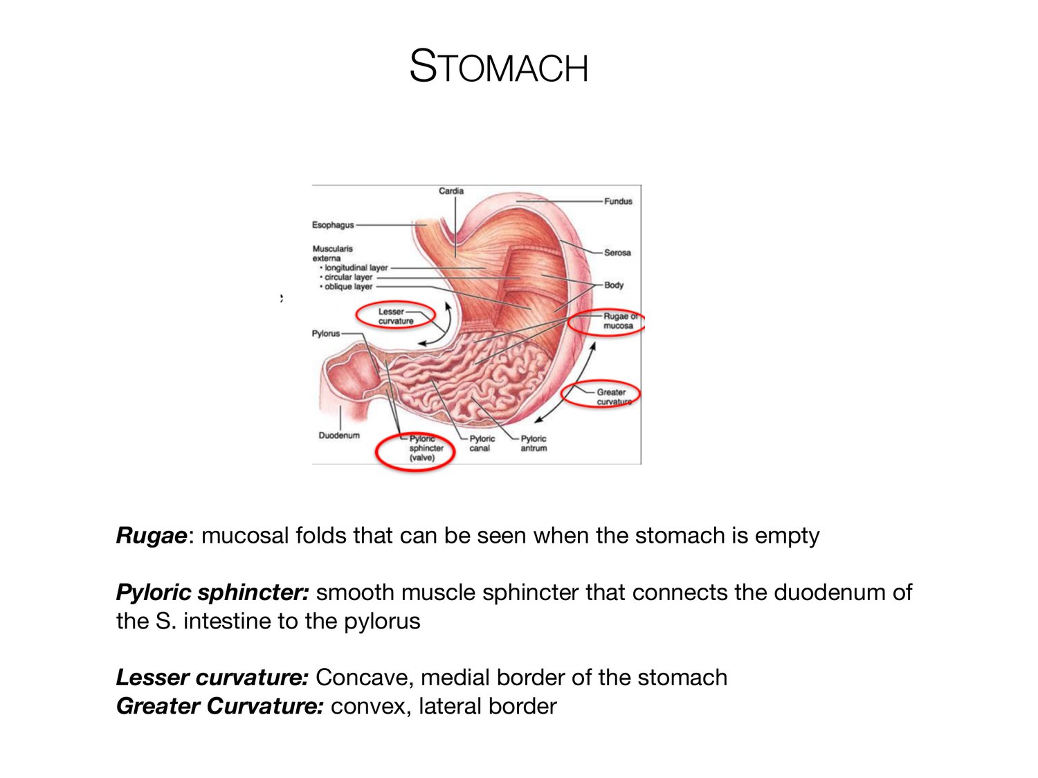
STOMACH Rugae: mucosal folds that can be seen when the stomach is empty Pyloric sphincter: smooth muscle sphincter that connects the duodenum of the S. intestine to the pylorus Lesser curvature: Concave, medial border of the stomach Greater Curvature: convex, lateral border
STOMACH-HISTOLOGY Histology of the Stomach (Deep to Superficial) Same basic layers as the rest of the GI tract; there are some modifications Surface of mucosa=simple columnar epithelium (aka: Surface Mucous Cells) Mucosa • There are columns of secretory cells aka gastric glands • Gastric pits: Several gastric glands that open into the bottom of narrow channels [Secretions from gastric glands flow in here and then to the lumen of the stomach. Lamina Propria=areolar CT Muscularis mucosae=smooth muscle tissue Submucosa Tissue: Areolar CT Tissue: 3 layers of smooth muscle tissue (esophagus has 2; S. & L. Intestines have 2) Muscularis • Outer longitudinal layer • Middle circular layer • Inner Oblique layer (limited to the body of the stomach) Serosa (visceral peritoneum) Tissue: simple squamous epithelium (mesothelium) & areolar CT At the lesser curvature: visceral peritoneum extends upward to the liver as lesser omentum Adventitia At the greater curvature: Visceral peritoneum extends downward as the greater omentum and drapes over the intestine
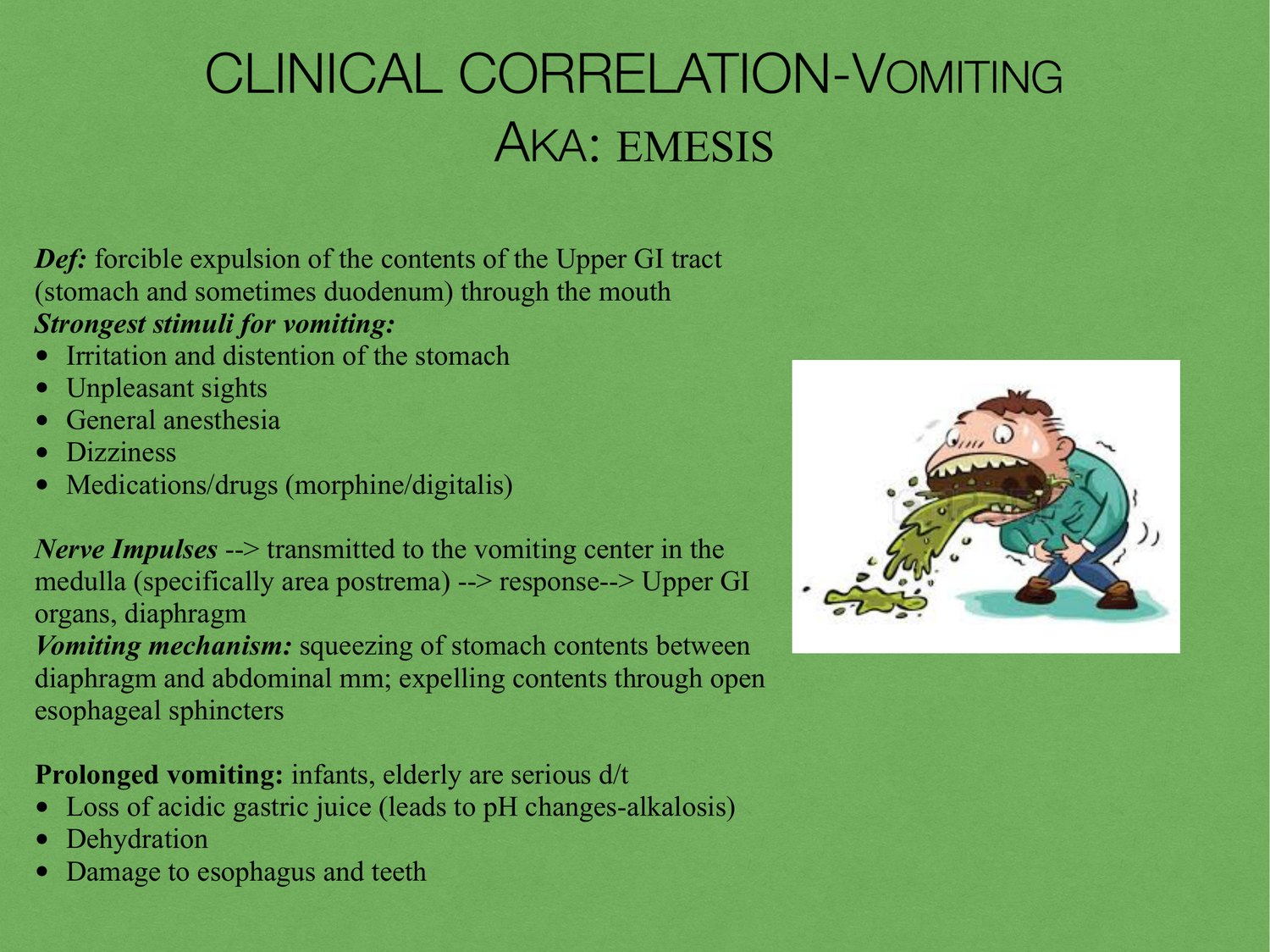
CLINICAL CORRELATION-VOMITING AKA: EMESIS Def: forcible expulsion of the contents of the Upper GI tract (stomach and sometimes duodenum) through the mouth Strongest stimuli for vomiting: • Irritation and distention of the stomach • Unpleasant sights • General anesthesia • Dizziness • Medications/drugs (morphine/digitalis) Nerve Impulses --> transmitted to the vomiting center in the medulla (specifically area postrema) --> response--> Upper GI organs, diaphragm Vomiting mechanism: squeezing of stomach contents between diaphragm and abdominal mm; expelling contents through open esophageal sphincters Prolonged vomiting: infants, elderly are serious d/t • Loss of acidic gastric juice (leads to pH changes-alkalosis) • Dehydration • Damage to esophagus and teeth
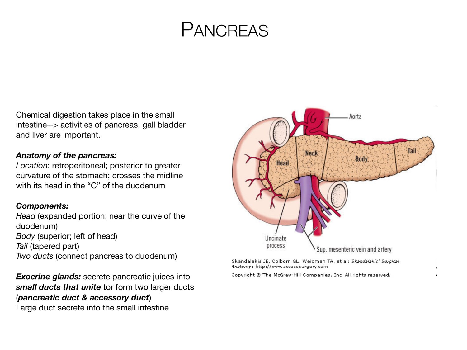
PANCREAS Chemical digestion takes place in the small intestine--> activities of pancreas, gall bladder and liver are important. Anatomy of the pancreas: Location: retroperitoneal; posterior to greater curvature of the stomach; crosses the midline with its head in the “C” of the duodenum Components: Head (expanded portion; near the curve of the duodenum) Body (superior; left of head) Tail (tapered part) Two ducts (connect pancreas to duodenum) Exocrine glands: secrete pancreatic juices into small ducts that unite tor form two larger ducts (pancreatic duct & accessory duct) Large duct secrete into the small intestine
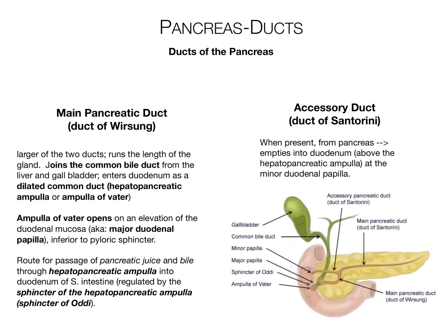
PANCREAS-DUCTS Ducts of the Pancreas Accessory Duct Main Pancreatic Duct (duct of Wirsung) (duct of Santorini) When present, from pancreas --> larger of the two ducts; runs the length of the empties into duodenum (above the gland. Joins the common bile duct from the hepatopancreatic ampulla) at the liver and gall bladder; enters duodenum as a minor duodenal papilla. dilated common duct (hepatopancreatic ampulla or ampulla of vater) Ampulla of vater opens on an elevation of the duodenal mucosa (aka: major duodenal papilla), inferior to pyloric sphincter. Route for passage of pancreatic juice and bile through hepatopancreatic ampulla into duodenum of S. intestine (regulated by the sphincter of the hepatopancreatic ampulla (sphincter of Oddi).
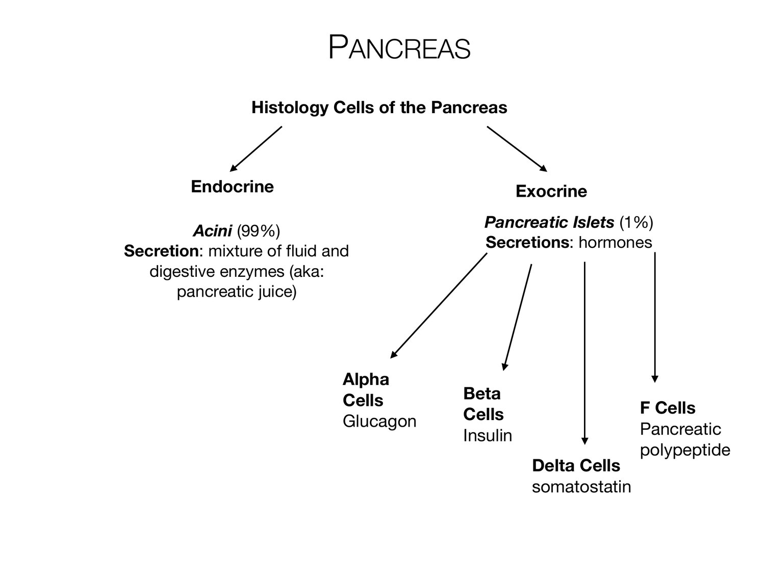
PANCREAS Histology Cells of the Pancreas Endocrine Exocrine Pancreatic Islets (1%) Acini (99%) Secretions: hormones Secretion: mixture of fluid and digestive enzymes (aka: pancreatic juice) Alpha Cells Beta Cells F Cells Glucagon Pancreatic Insulin polypeptide Delta Cells somatostatin
PANCREAS-BLOOD SUPPLY Venous Arterial Drains into splenic and superior Branches of the celiac trunk mesenteric veins; merge to Superior mesenteric artery: form the hepatic portal system.
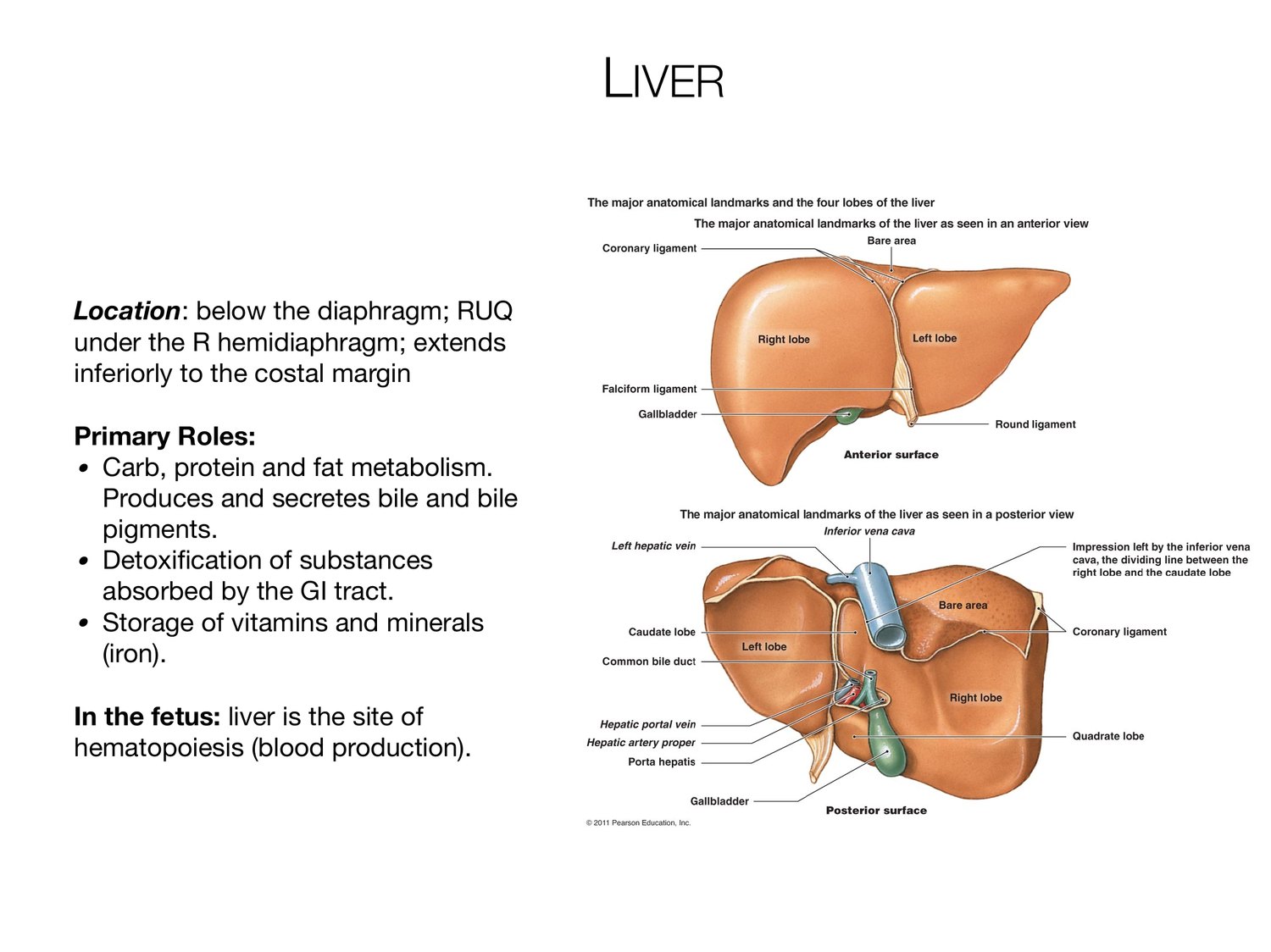
LIVER Location: below the diaphragm; RUQ under the R hemidiaphragm; extends inferiorly to the costal margin Primary Roles: • Carb, protein and fat metabolism. Produces and secretes bile and bile pigments. • Detoxification of substances absorbed by the GI tract. • Storage of vitamins and minerals (iron). In the fetus: liver is the site of hematopoiesis (blood production).
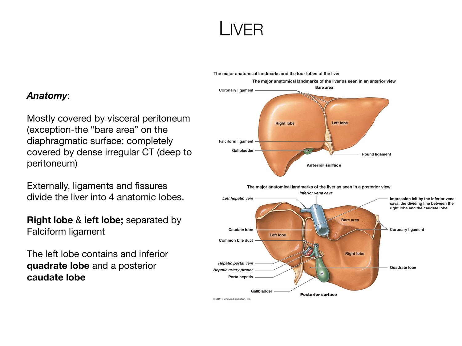
LIVER Anatomy: Mostly covered by visceral peritoneum (exception-the “bare area” on the diaphragmatic surface; completely covered by dense irregular CT (deep to peritoneum) Externally, ligaments and fissures divide the liver into 4 anatomic lobes. Right lobe & left lobe; separated by Falciform ligament The left lobe contains and inferior quadrate lobe and a posterior caudate lobe
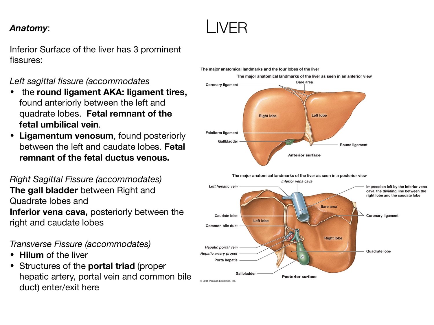
Anatomy: LIVER Inferior Surface of the liver has 3 prominent fissures: Left sagittal fissure (accommodates • the round ligament AKA: ligament tires, found anteriorly between the left and quadrate lobes. Fetal remnant of the fetal umbilical vein. • Ligamentum venosum, found posteriorly between the left and caudate lobes. Fetal remnant of the fetal ductus venous. Right Sagittal Fissure (accommodates) The gall bladder between Right and Quadrate lobes and Inferior vena cava, posteriorly between the right and caudate lobes Transverse Fissure (accommodates) • Hilum of the liver • Structures of the portal triad (proper hepatic artery, portal vein and common bile duct) enter/exit here
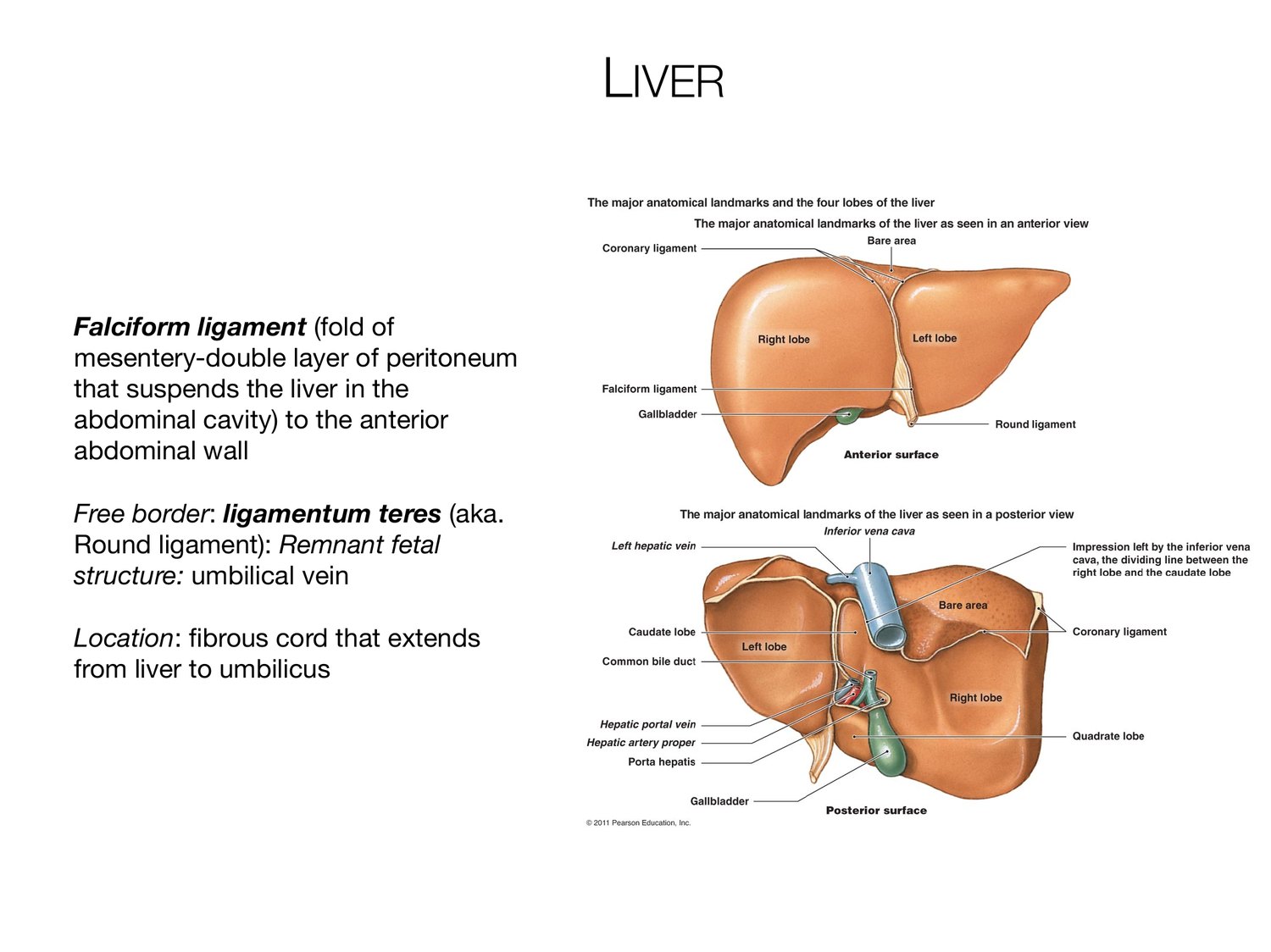
LIVER Falciform ligament (fold of mesentery-double layer of peritoneum that suspends the liver in the abdominal cavity) to the anterior abdominal wall Free border: ligamentum teres (aka. Round ligament): Remnant fetal structure: umbilical vein Location: fibrous cord that extends from liver to umbilicus
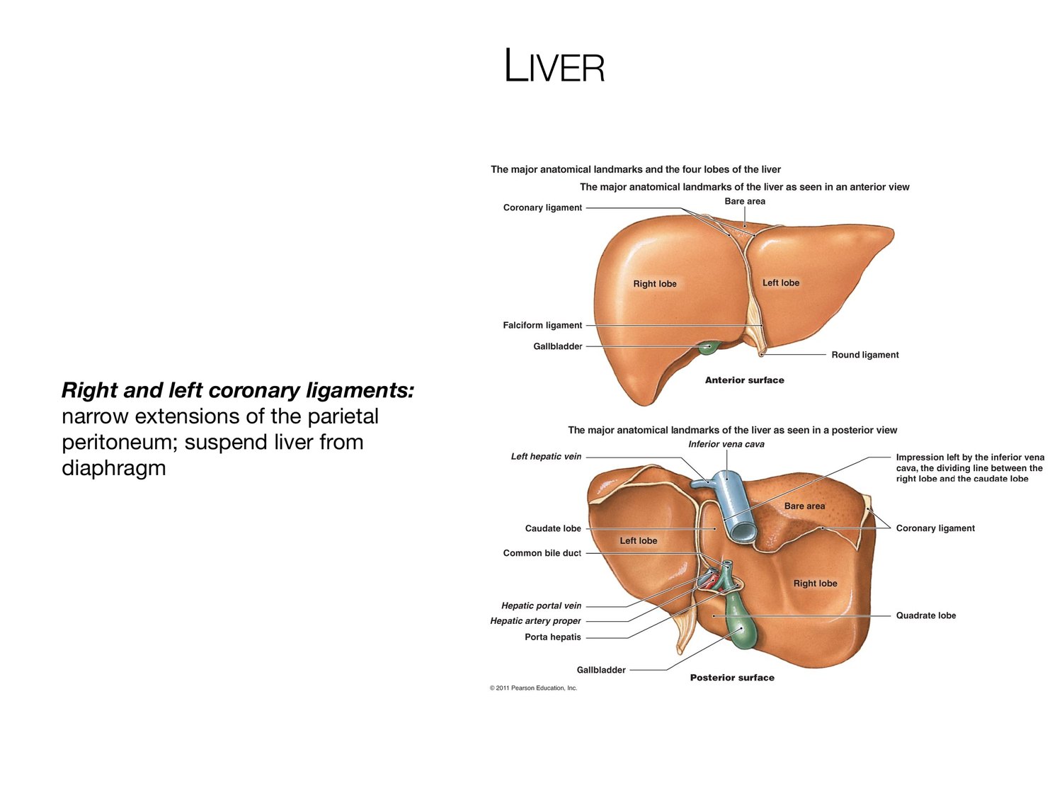
LIVER Right and left coronary ligaments: narrow extensions of the parietal peritoneum; suspend liver from diaphragm
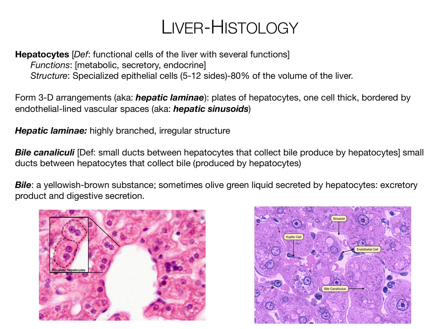
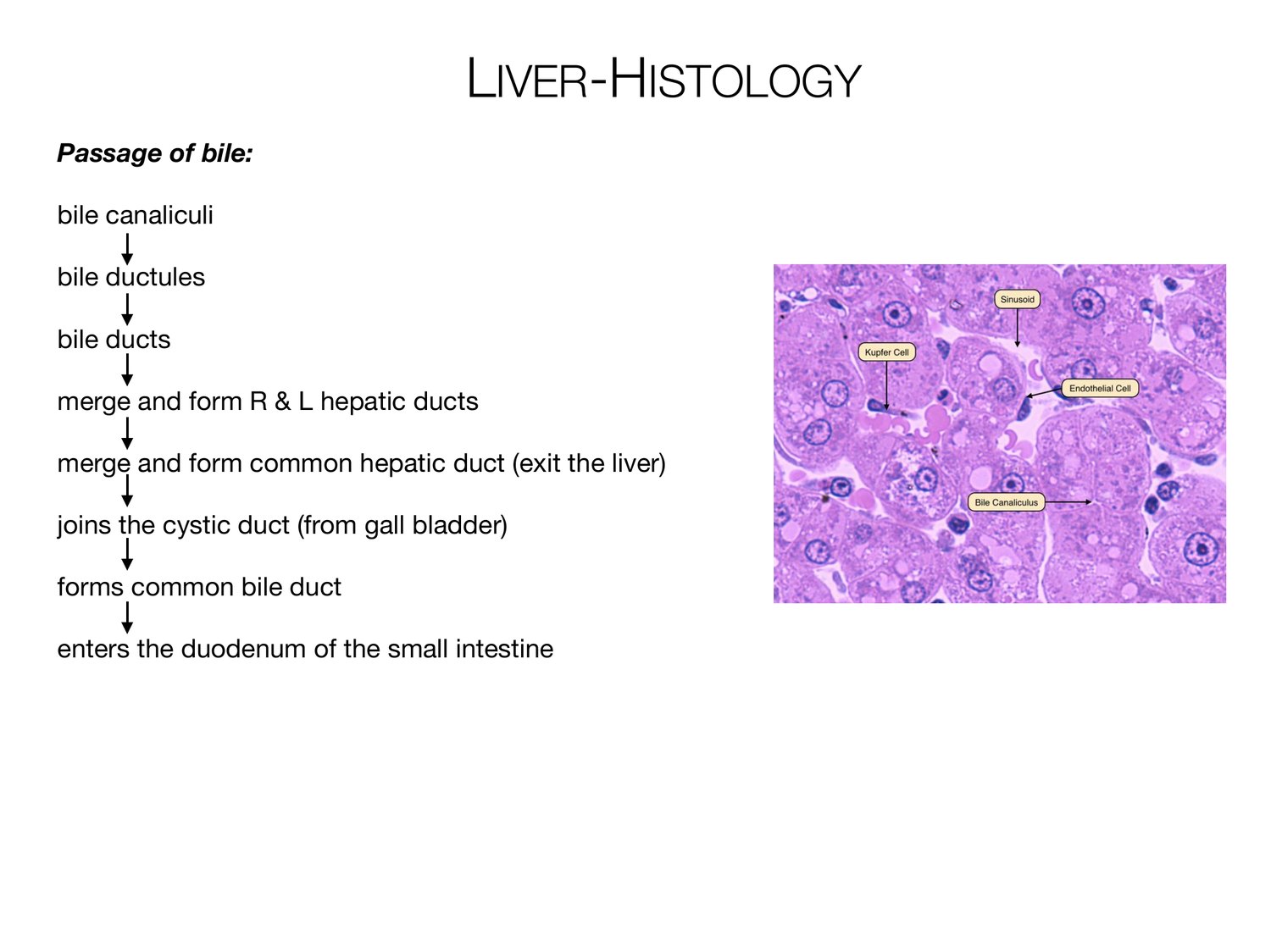
LIVER-HISTOLOGY Hepatocytes [Def: functional cells of the liver with several functions] Functions: [metabolic, secretory, endocrine] Structure: Specialized epithelial cells (5-12 sides)-80% of the volume of the liver. Form 3-D arrangements (aka: hepatic laminae): plates of hepatocytes, one cell thick, bordered by endothelial-lined vascular spaces (aka: hepatic sinusoids) Hepatic laminae: highly branched, irregular structure Bile canaliculi [Def: small ducts between hepatocytes that collect bile produce by hepatocytes] small ducts between hepatocytes that collect bile (produced by hepatocytes) Bile: a yellowish-brown substance; sometimes olive green liquid secreted by hepatocytes: excretory product and digestive secretion.
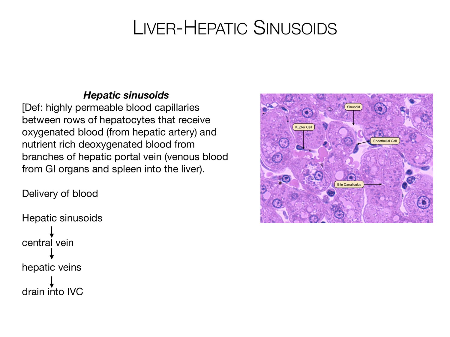
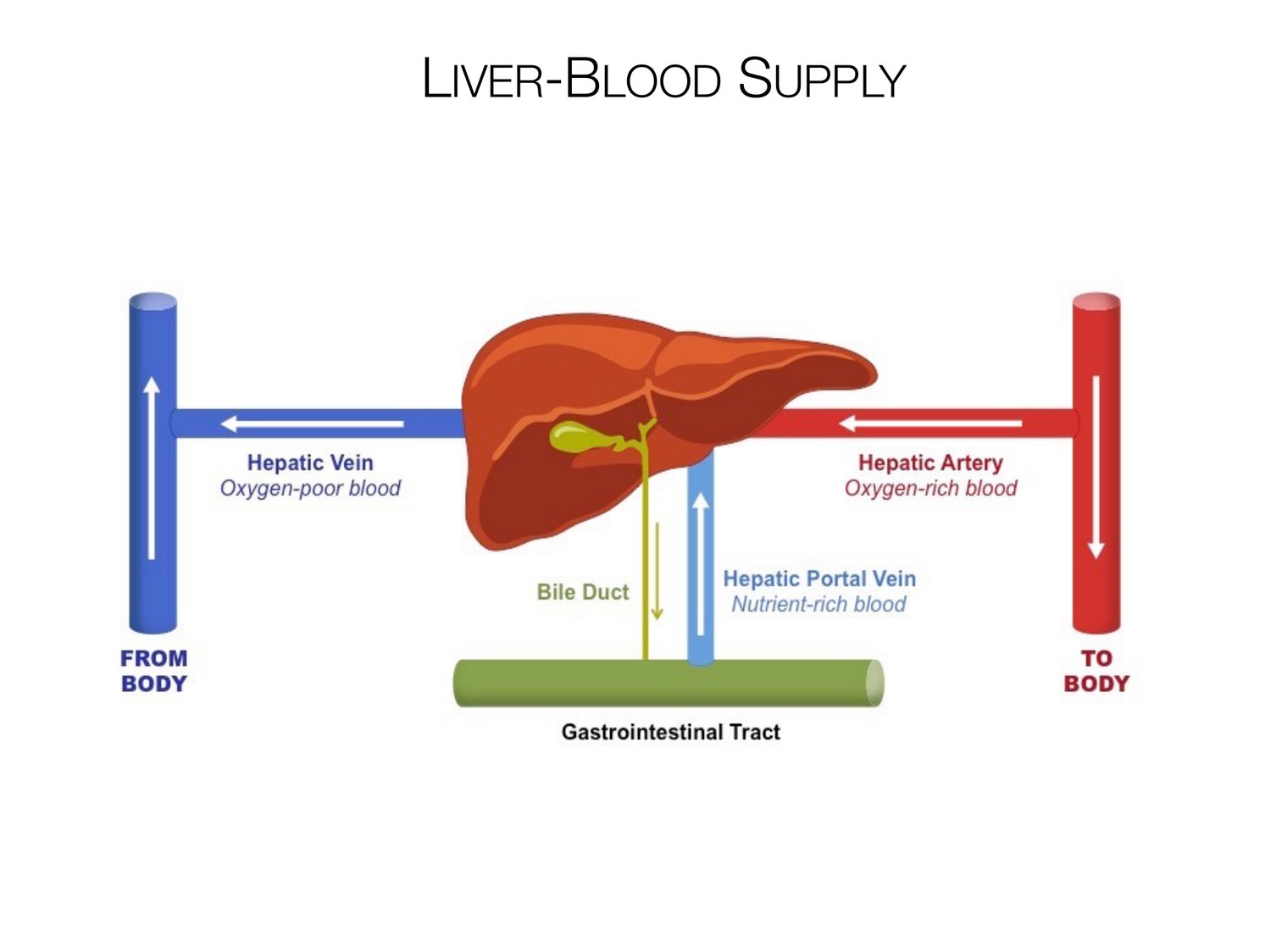
LIVER-HEPATIC SINUSOIDS Hepatic sinusoids [Def: highly permeable blood capillaries between rows of hepatocytes that receive oxygenated blood (from hepatic artery) and nutrient rich deoxygenated blood from branches of hepatic portal vein (venous blood from GI organs and spleen into the liver). Delivery of blood Hepatic sinusoids central vein hepatic veins drain into IVC
LIVER-HISTOLOGY Passage of bile: bile canaliculi bile ductules bile ducts merge and form R & L hepatic ducts merge and form common hepatic duct (exit the liver) joins the cystic duct (from gall bladder) forms common bile duct enters the duodenum of the small intestine
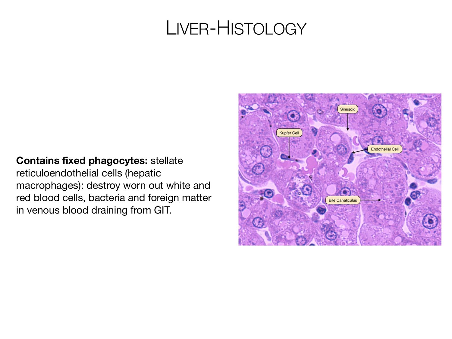
LIVER-HISTOLOGY Contains fixed phagocytes: stellate reticuloendothelial cells (hepatic macrophages): destroy worn out white and red blood cells, bacteria and foreign matter in venous blood draining from GIT.
LIVER-BLOOD SUPPLY
LIVER-BLOOD SUPPLY Arterial Venous Dual Blood Supply: Portal Vein & Hepatic Artery Right, left and intermediate hepatic Proper. Both divide into primary and secondary veins drain segments and dump branches. into the inferior vena cava. • Portal vein: carries nutrient rich blood from the digestive tract. (75-85% of blood volume carried to liver via this structure). (Deoxygenated blood with newly absorbed nutrients from GIT) • Hepatic artery proper: supplied by celiac trunk via common hepatic artery. (20-25% of blood volume to the liver) (Oxygenated blood) Hepatic artery + Hepatic portal vein --> hepatic sinusoids --> central vein --> hepatic vein --: IVC --> R. atrium Due to the significant amount of blood that flow through here, this is a very common site for cancer metastasis. Internal branching of intrahepatic blood vessels divides liver into 8 functional segments. Blood supply facilitates the resection of individual diseased segments.
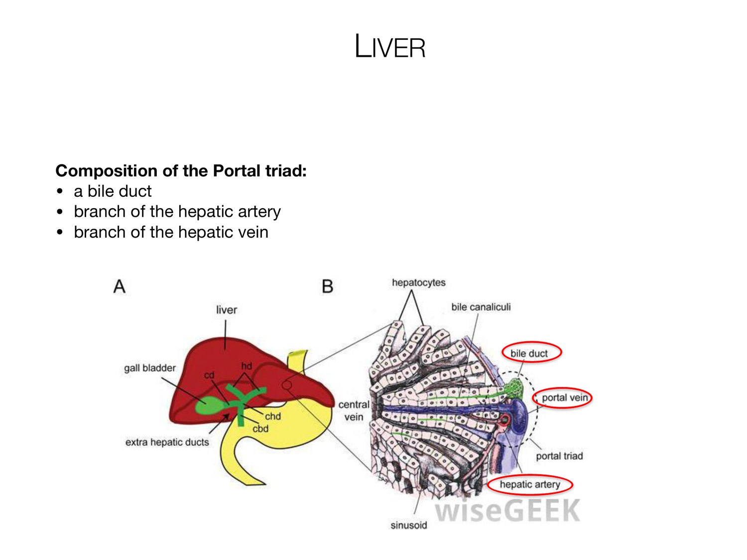
LIVER Composition of the Portal triad: • a bile duct • branch of the hepatic artery • branch of the hepatic vein
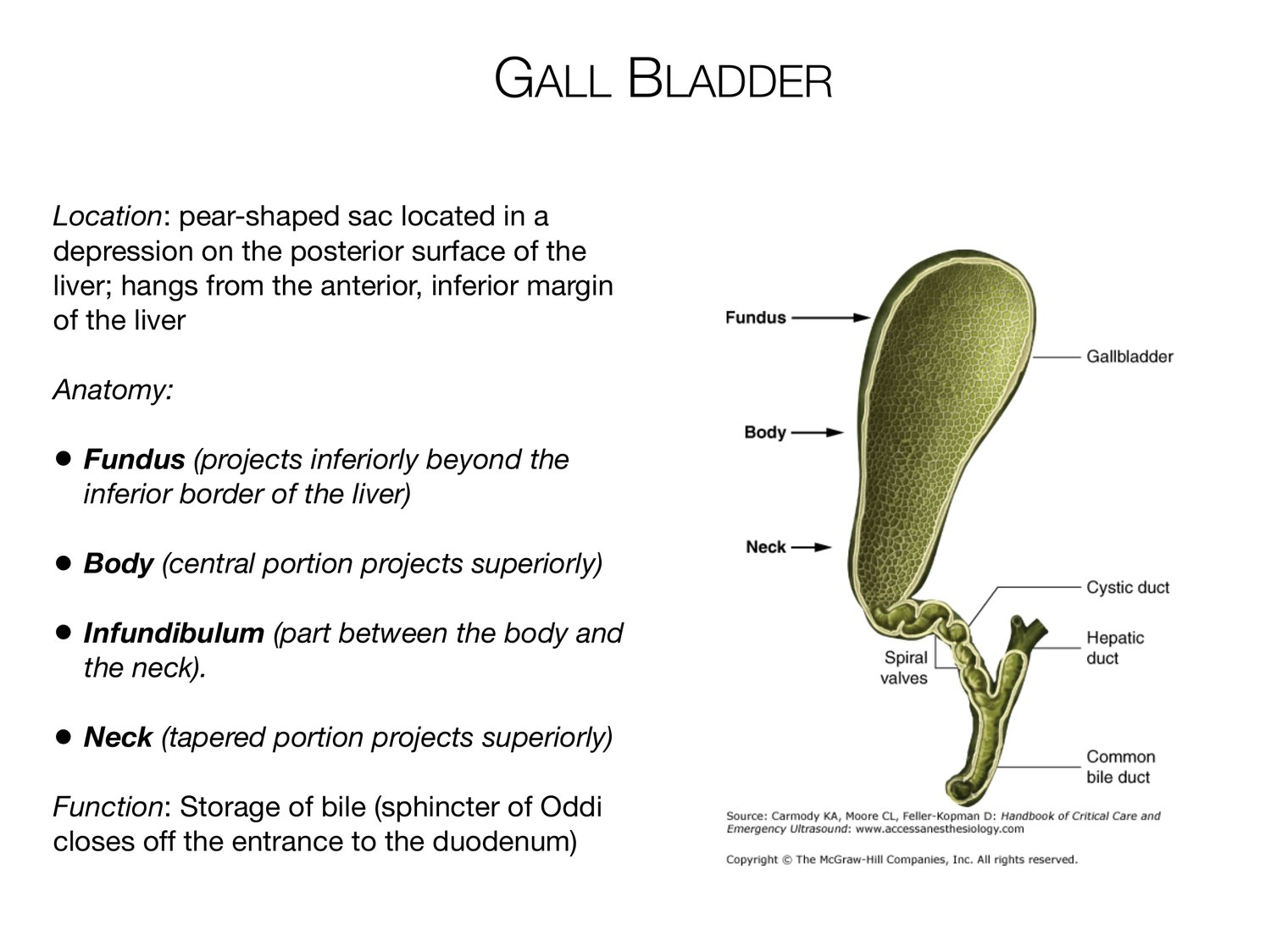
GALL BLADDER Location: pear-shaped sac located in a depression on the posterior surface of the liver; hangs from the anterior, inferior margin of the liver Anatomy: • Fundus (projects inferiorly beyond the inferior border of the liver) • Body (central portion projects superiorly) • Infundibulum (part between the body and the neck). • Neck (tapered portion projects superiorly) Function: Storage of bile (sphincter of Oddi closes off the entrance to the duodenum)
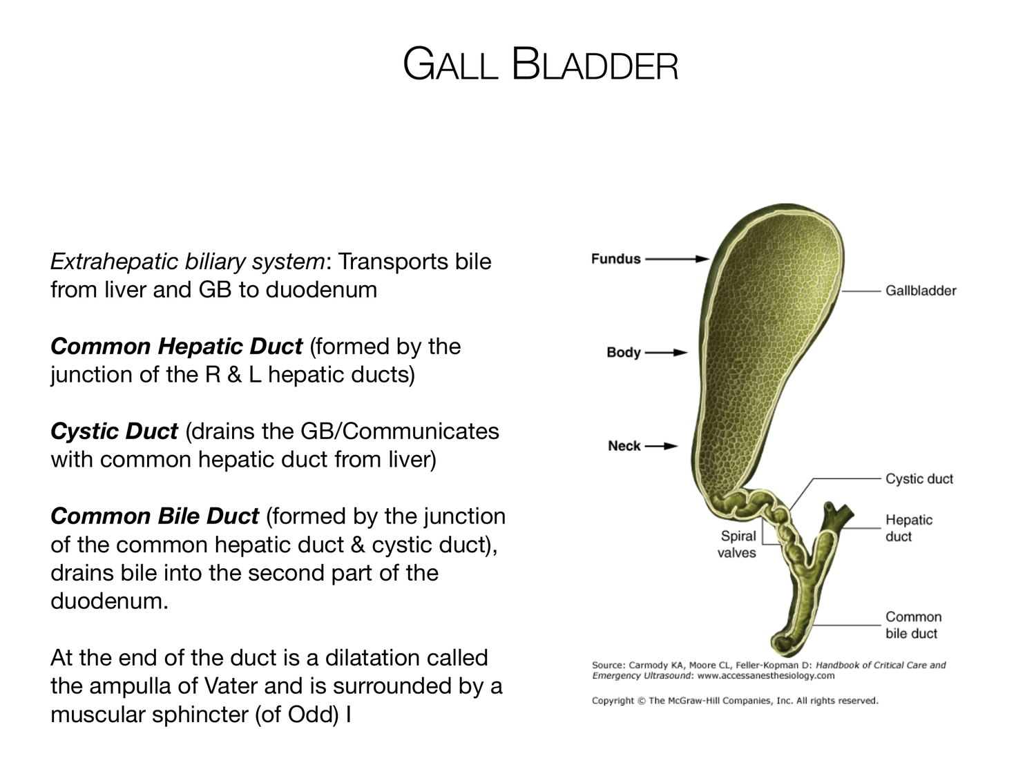
GALL BLADDER Extrahepatic biliary system: Transports bile from liver and GB to duodenum Common Hepatic Duct (formed by the junction of the R & L hepatic ducts) Cystic Duct (drains the GB/Communicates with common hepatic duct from liver) Common Bile Duct (formed by the junction of the common hepatic duct & cystic duct), drains bile into the second part of the duodenum. At the end of the duct is a dilatation called the ampulla of Vater and is surrounded by a muscular sphincter (of Odd) I
SMALL INTESTINE Three Regions of the Small Intestine 1. Duodenum (shortest; retroperitoneal; begins: pyloric sphincter! ends at merging with jejunum; C shaped; 12”) 2. Jejunum (means empty-how it is found at death; 3 ft. long extends to ileum) 3. Ileum (longest; terminal part of small intestine; 6 ft. long, joins the large intestine at the ileocecal sphincter (valve). Ileocecal sphincter (valve): smooth muscle at the junction of the large intestine and the terminal part of the ileum
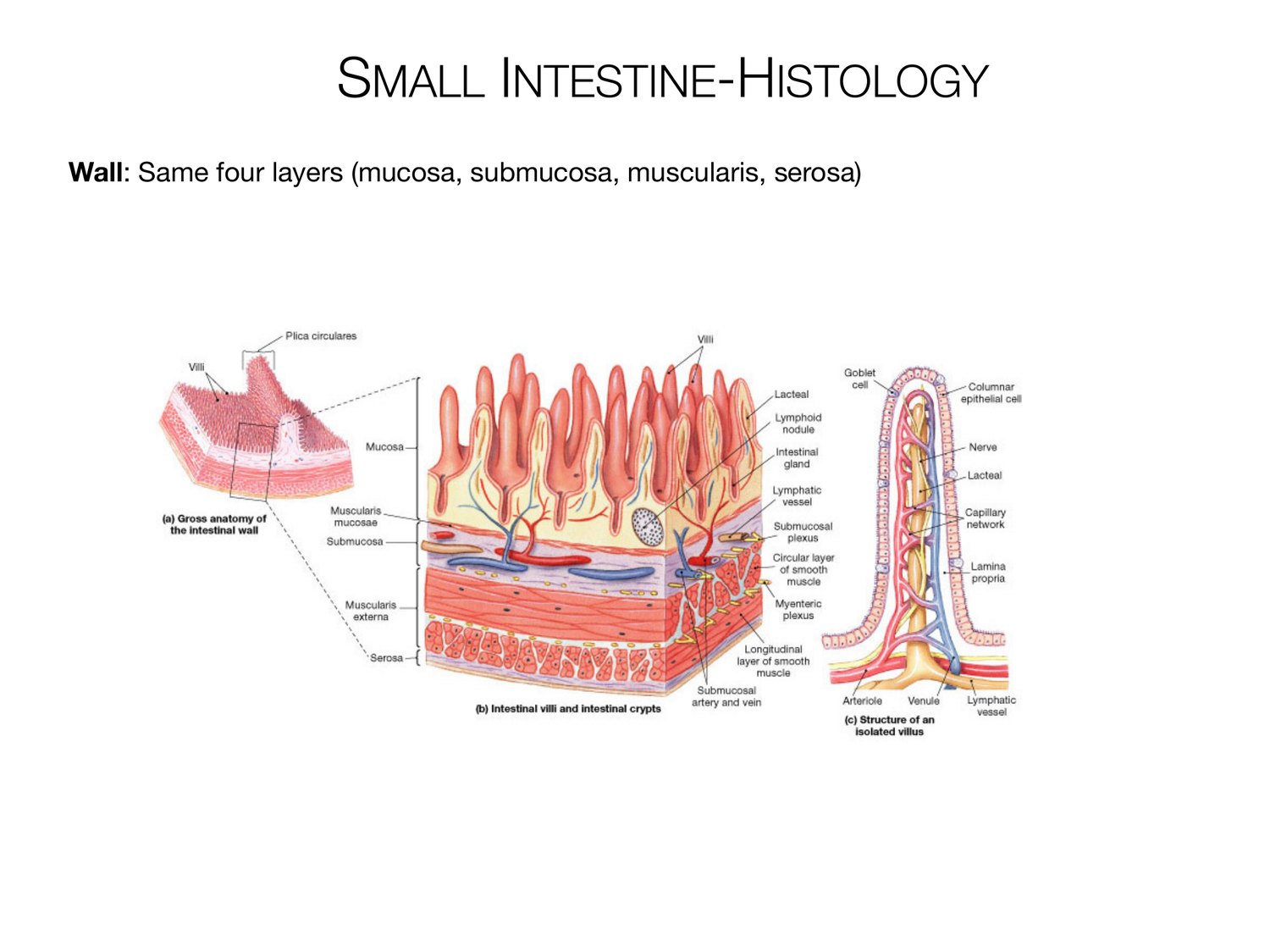
SMALL INTESTINE-HISTOLOGY Wall: Same four layers (mucosa, submucosa, muscularis, serosa)
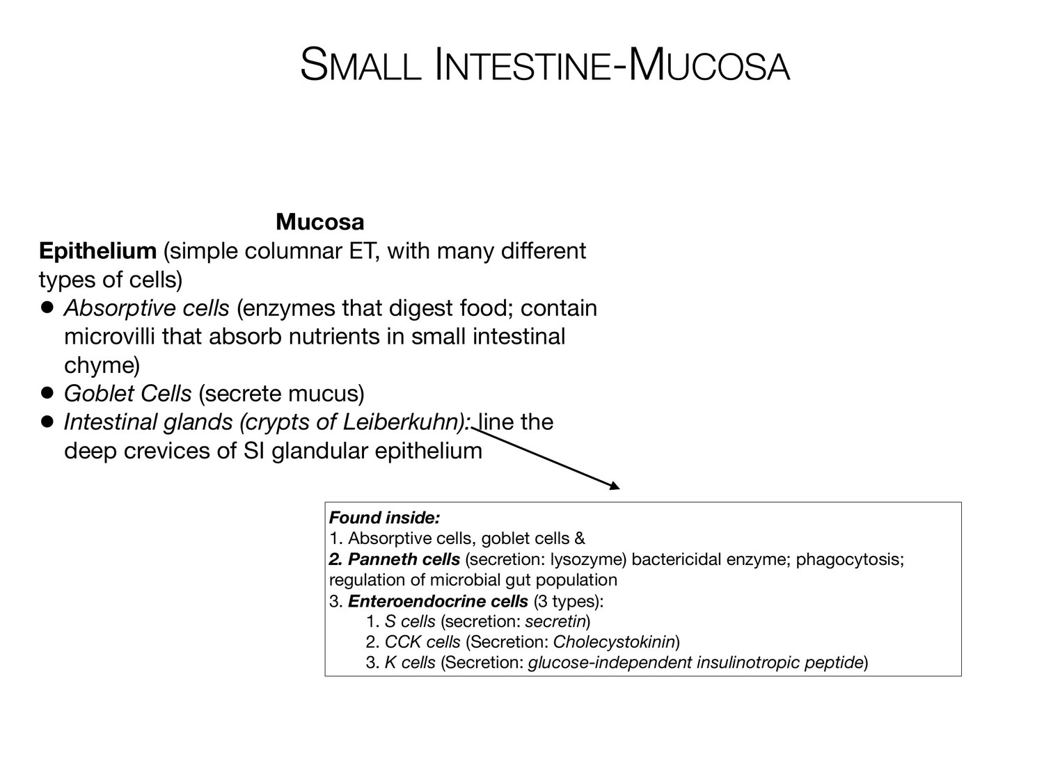
SMALL INTESTINE-MUCOSA Mucosa Epithelium (simple columnar ET, with many different types of cells) • Absorptive cells (enzymes that digest food; contain microvilli that absorb nutrients in small intestinal chyme) • Goblet Cells (secrete mucus) • Intestinal glands (crypts of Leiberkuhn): line the deep crevices of SI glandular epithelium Found inside: 1. Absorptive cells, goblet cells & 2. Panneth cells (secretion: lysozyme) bactericidal enzyme; phagocytosis; regulation of microbial gut population 3. Enteroendocrine cells (3 types): 1. S cells (secretion: secretin) 2. CCK cells (Secretion: Cholecystokinin) 3. K cells (Secretion: glucose-independent insulinotropic peptide)
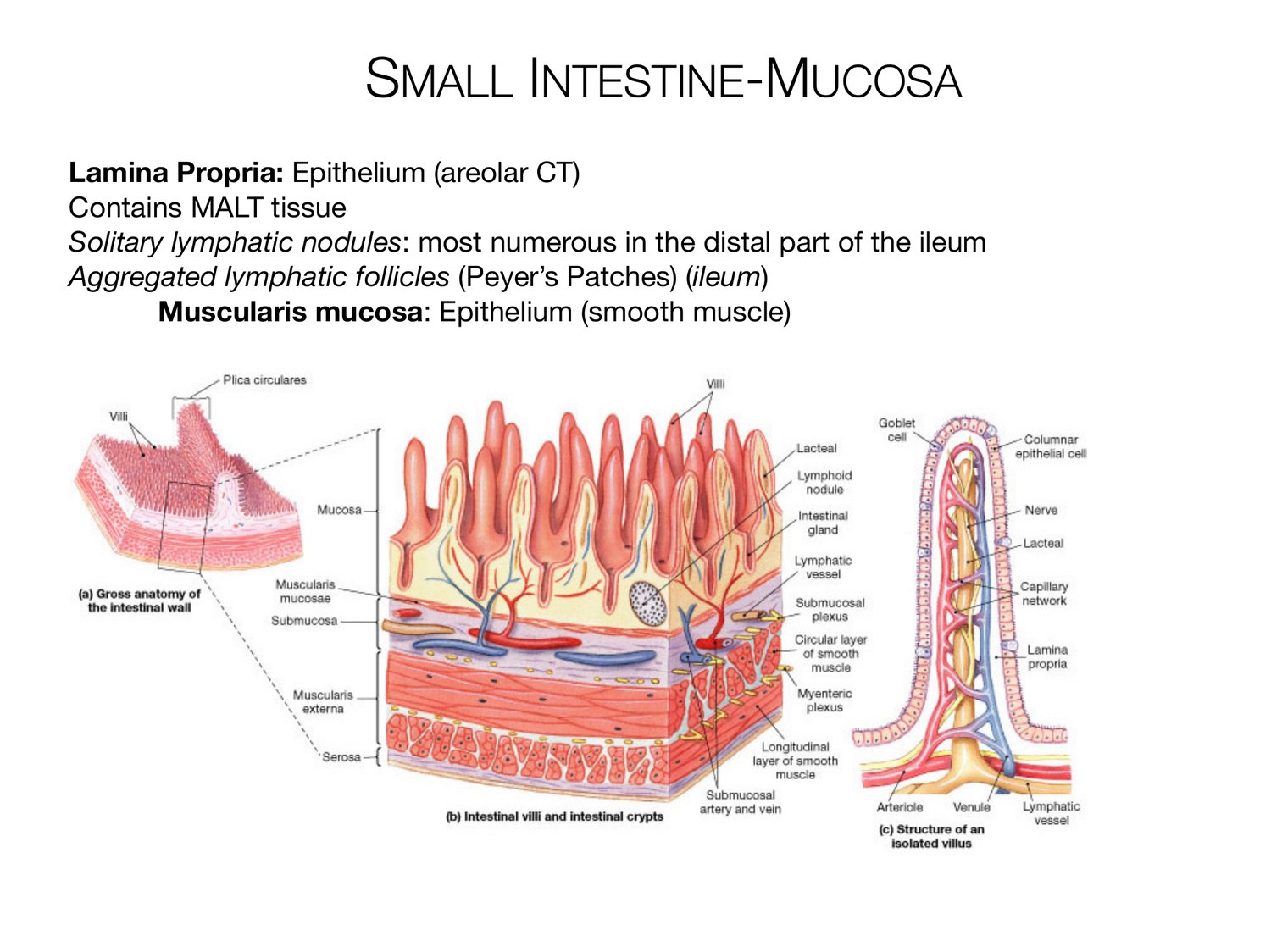
SMALL INTESTINE-MUCOSA Lamina Propria: Epithelium (areolar CT) Contains MALT tissue Solitary lymphatic nodules: most numerous in the distal part of the ileum Aggregated lymphatic follicles (Peyer’s Patches) (ileum) Muscularis mucosa: Epithelium (smooth muscle)
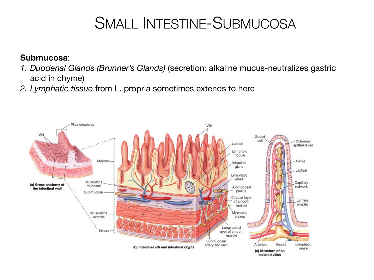
SMALL INTESTINE-SUBMUCOSA Submucosa: 1. Duodenal Glands (Brunner’s Glands) (secretion: alkaline mucus-neutralizes gastric acid in chyme) 2. Lymphatic tissue from L. propria sometimes extends to here
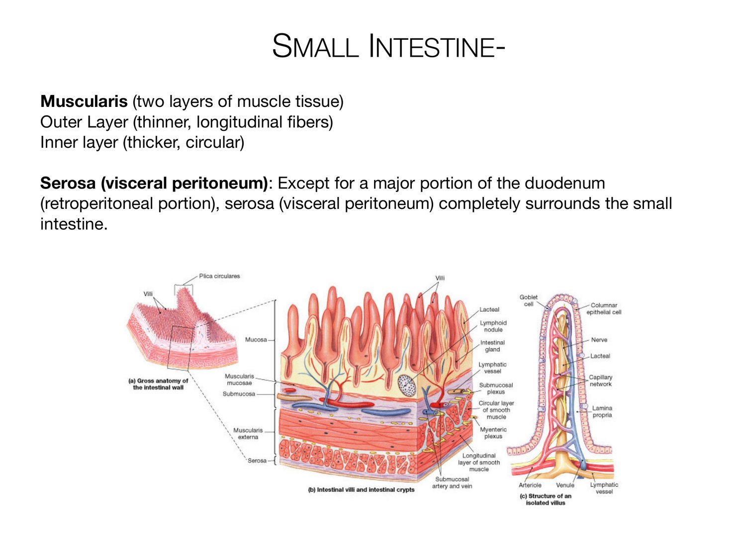
SMALL INTESTINE- Muscularis (two layers of muscle tissue) Outer Layer (thinner, longitudinal fibers) Inner layer (thicker, circular) Serosa (visceral peritoneum): Except for a major portion of the duodenum (retroperitoneal portion), serosa (visceral peritoneum) completely surrounds the small intestine.
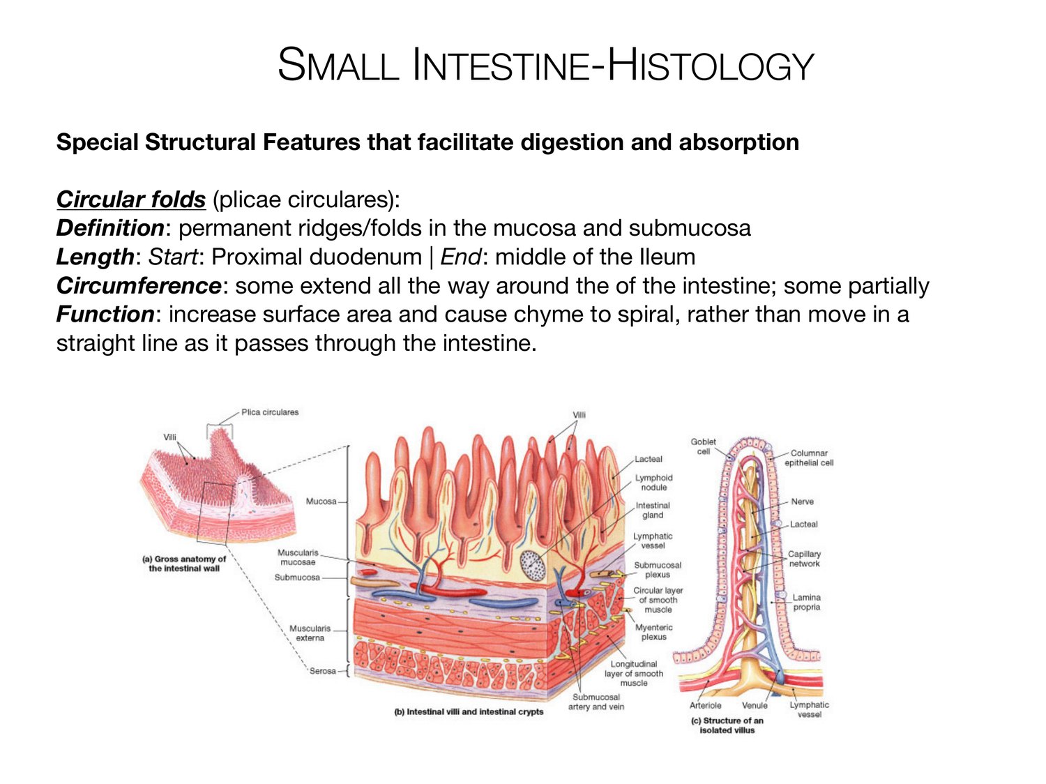
SMALL INTESTINE-HISTOLOGY Special Structural Features that facilitate digestion and absorption Circular folds (plicae circulares): Definition: permanent ridges/folds in the mucosa and submucosa Length: Start: Proximal duodenum | End: middle of the Ileum Circumference: some extend all the way around the of the intestine; some partially Function: increase surface area and cause chyme to spiral, rather than move in a straight line as it passes through the intestine.
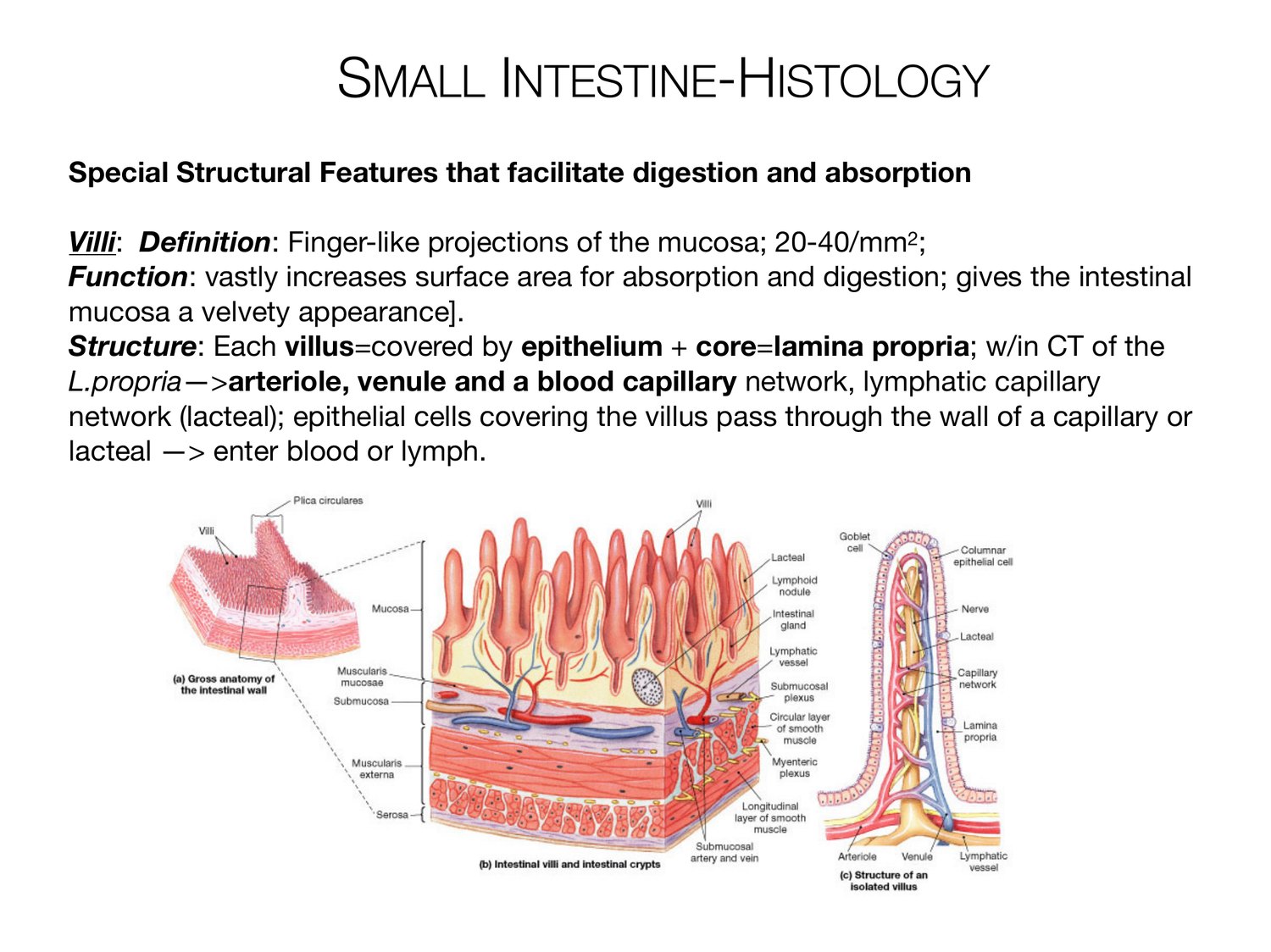
SMALL INTESTINE-HISTOLOGY Special Structural Features that facilitate digestion and absorption Villi: Definition: Finger-like projections of the mucosa; 20-40/mm2; Function: vastly increases surface area for absorption and digestion; gives the intestinal mucosa a velvety appearance]. Structure: Each villus=covered by epithelium + core=lamina propria; w/in CT of the L.propria->arteriole, venule and a blood capillary network, lymphatic capillary network (lacteal); epithelial cells covering the villus pass through the wall of a capillary or lacteal -> enter blood or lymph.
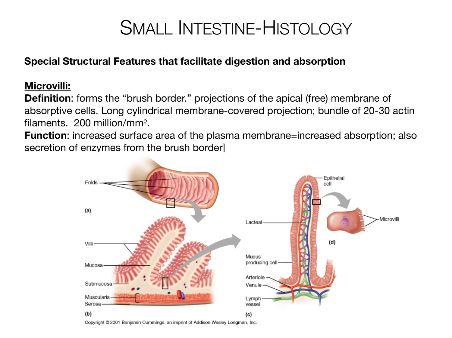
SMALL INTESTINE-HISTOLOGY Special Structural Features that facilitate digestion and absorption Microvilli: Definition: forms the “brush border.” projections of the apical (free) membrane of absorptive cells. Long cylindrical membrane-covered projection; bundle of 20-30 actin filaments. 200 million/mm2. Function: increased surface area of the plasma membrane=increased absorption; also secretion of enzymes from the brush border]
LARGE INTESTINE Large Intestine Def: terminal part of the GI tract Functions: 1. Haustral churning, peristalsis and mass peristalsis drive contents of colon into rectum 2. Bacteria in Lg. intestine convert proteins into AA; break down AA and produce some B vitamins and Vitamin K 3. Absorption of some water, ions and vitamins 4. Formation of feces 5. Defecation (emptying of the rectum)
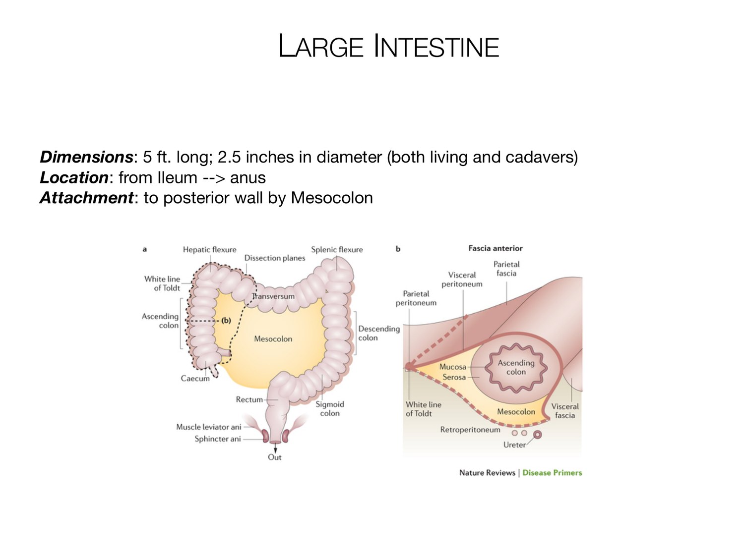
LARGE INTESTINE Dimensions: 5 ft. long; 2.5 inches in diameter (both living and cadavers) Location: from Ileum --> anus Attachment: to posterior wall by Mesocolon
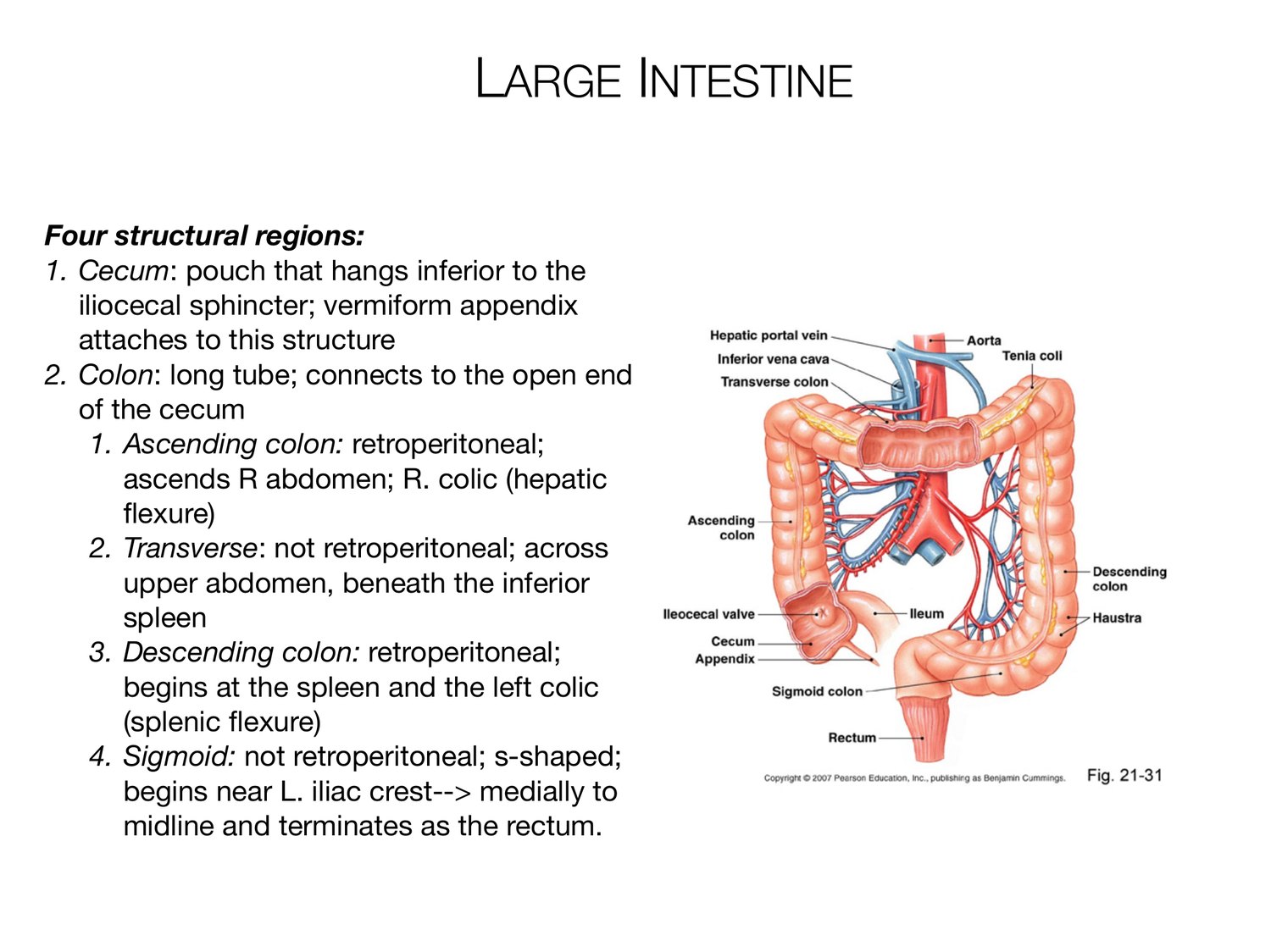
LARGE INTESTINE Four structural regions: 1. Cecum: pouch that hangs inferior to the iliocecal sphincter; vermiform appendix attaches to this structure 2. Colon: long tube; connects to the open end of the cecum 1. Ascending colon: retroperitoneal; ascends R abdomen; R. colic (hepatic flexure) 2. Transverse: not retroperitoneal; across upper abdomen, beneath the inferior spleen 3. Descending colon: retroperitoneal; begins at the spleen and the left colic (splenic flexure) 4. Sigmoid: not retroperitoneal; s-shaped; begins near L. iliac crest--> medially to midline and terminates as the rectum.
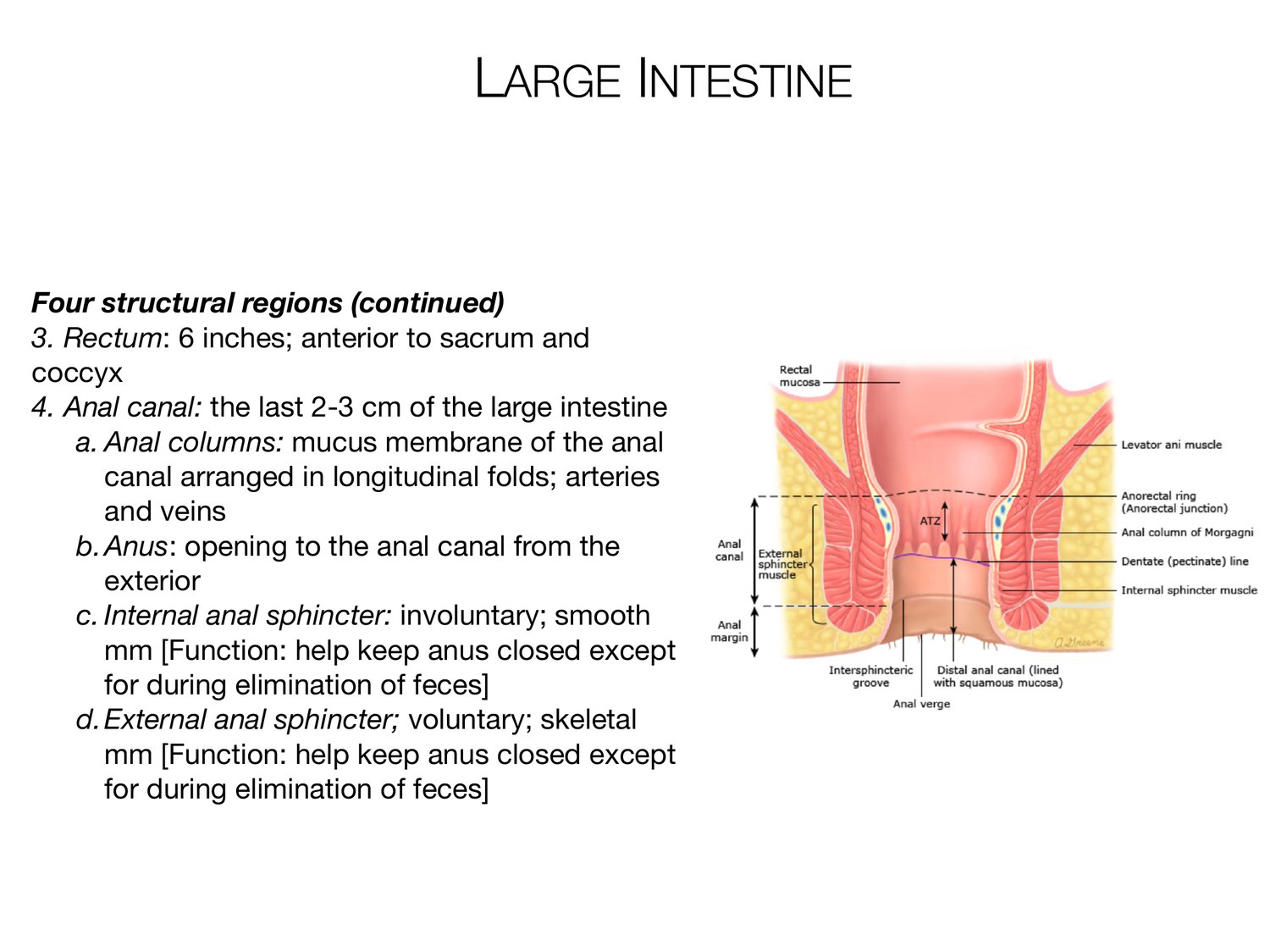
LARGE INTESTINE Four structural regions (continued) 3. Rectum: 6 inches; anterior to sacrum and coccyx 4. Anal canal: the last 2-3 cm of the large intestine a. Anal columns: mucus membrane of the anal canal arranged in longitudinal folds; arteries and veins b. Anus: opening to the anal canal from the exterior c. Internal anal sphincter: involuntary; smooth mm [Function: help keep anus closed except for during elimination of feces] d. External anal sphincter; voluntary; skeletal mm [Function: help keep anus closed except for during elimination of feces]
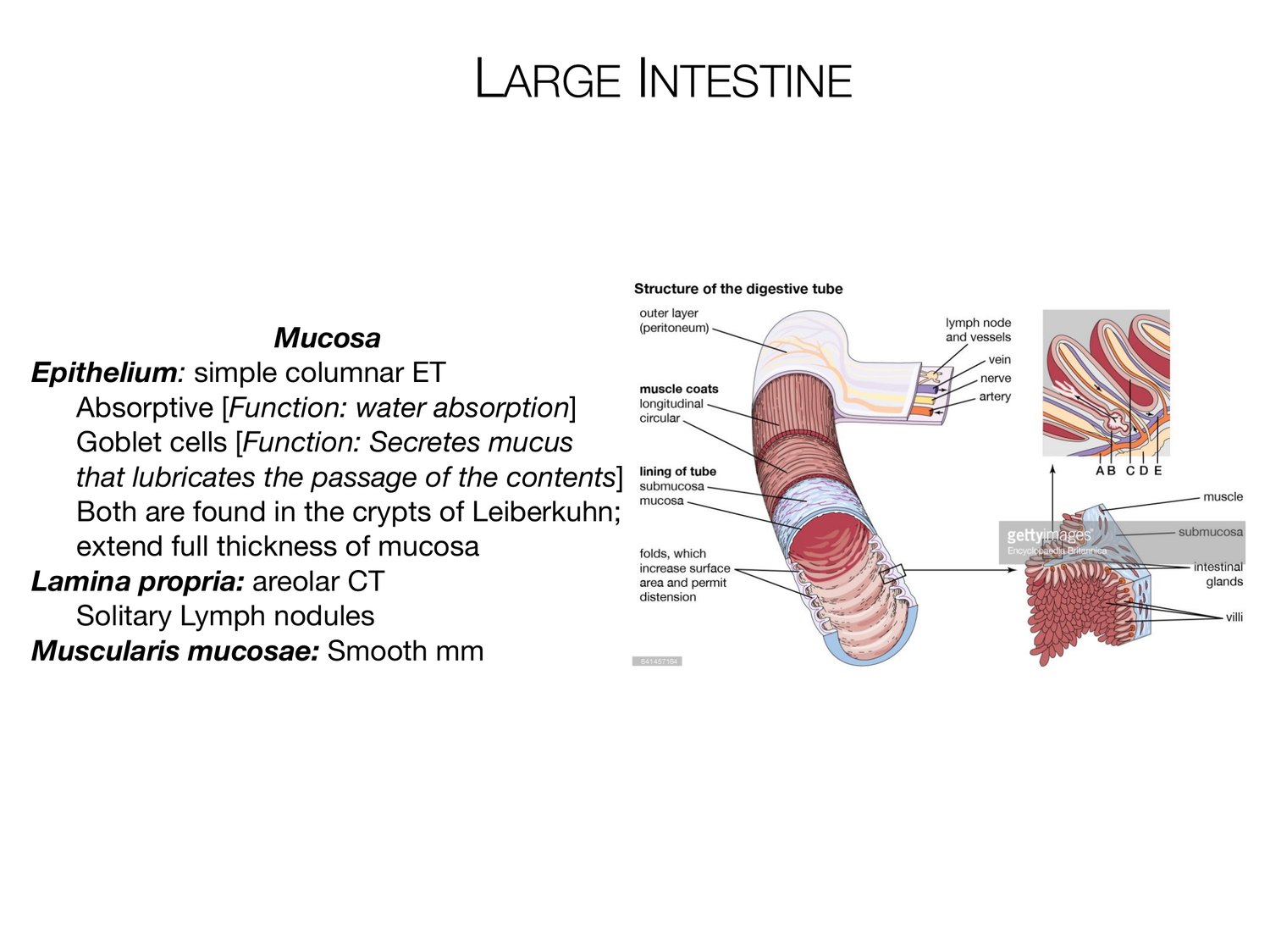
LARGE INTESTINE Mucosa Epithelium: simple columnar ET Absorptive [Function: water absorption] Goblet cells [Function: Secretes mucus that lubricates the passage of the contents] Both are found in the crypts of Leiberkuhn; extend full thickness of mucosa Lamina propria: areolar CT Solitary Lymph nodules Muscularis mucosae: Smooth mm
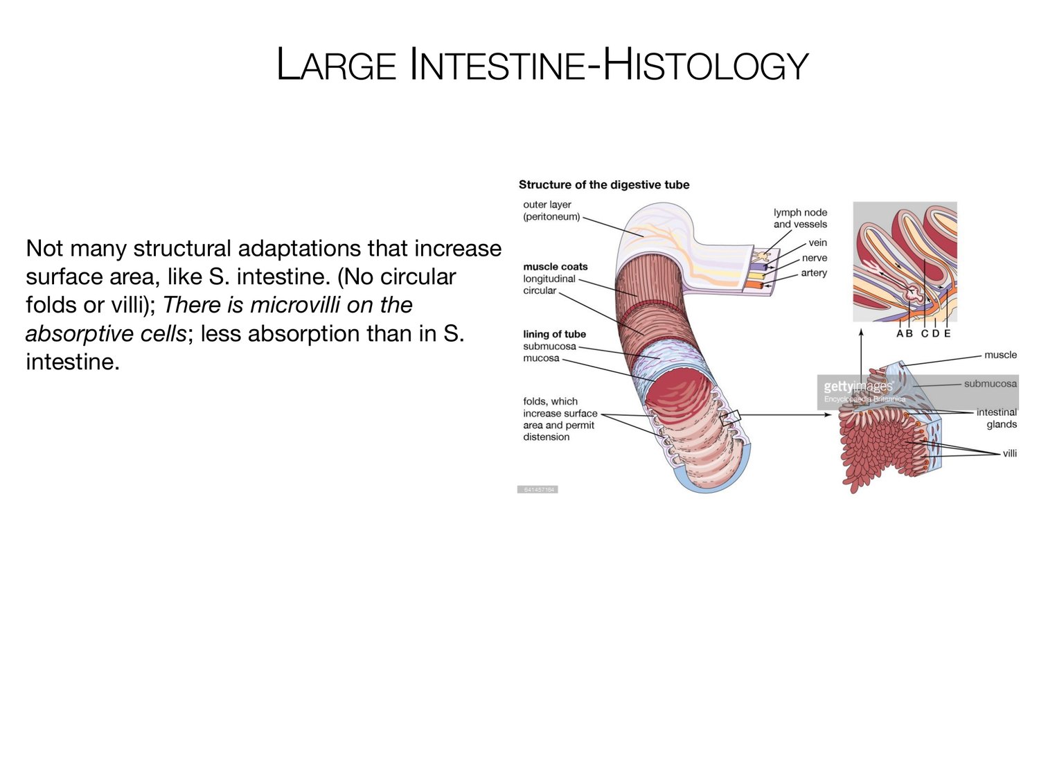
LARGE INTESTINE-HISTOLOGY Not many structural adaptations that increase surface area, like S. intestine. (No circular folds or villi); There is microvilli on the absorptive cells; less absorption than in S. intestine.
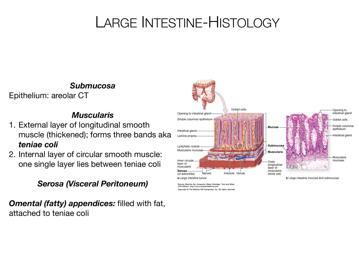
LARGE INTESTINE-HISTOLOGY Submucosa Epithelium: areolar CT Muscularis 1. External layer of longitudinal smooth muscle (thickened); forms three bands aka teniae coli 2. Internal layer of circular smooth muscle: one single layer lies between teniae coli Serosa (Visceral Peritoneum) Omental (fatty) appendices: filled with fat, attached to teniae coli
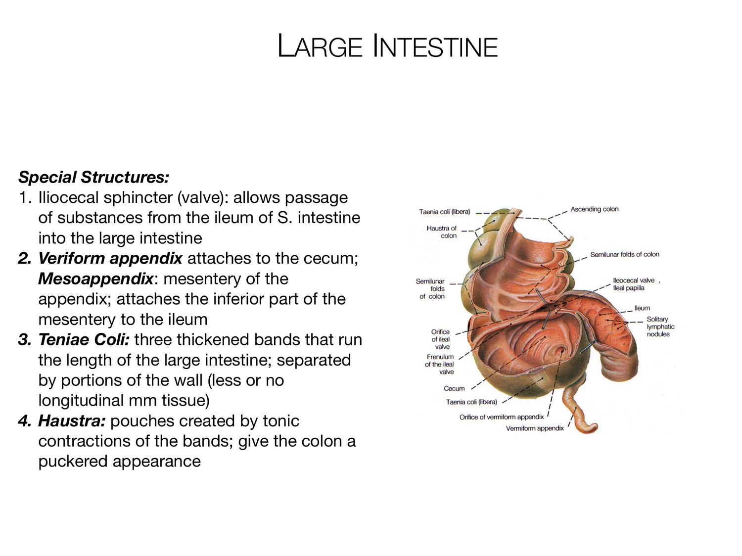
LARGE INTESTINE Special Structures: 1. Iliocecal sphincter (valve): allows passage of substances from the ileum of S. intestine into the large intestine 2. Veriform appendix attaches to the cecum; Mesoappendix: mesentery of the appendix; attaches the inferior part of the mesentery to the ileum 3. Teniae Coli: three thickened bands that run the length of the large intestine; separated by portions of the wall (less or no longitudinal mm tissue) 4. Haustra: pouches created by tonic contractions of the bands; give the colon a puckered appearance
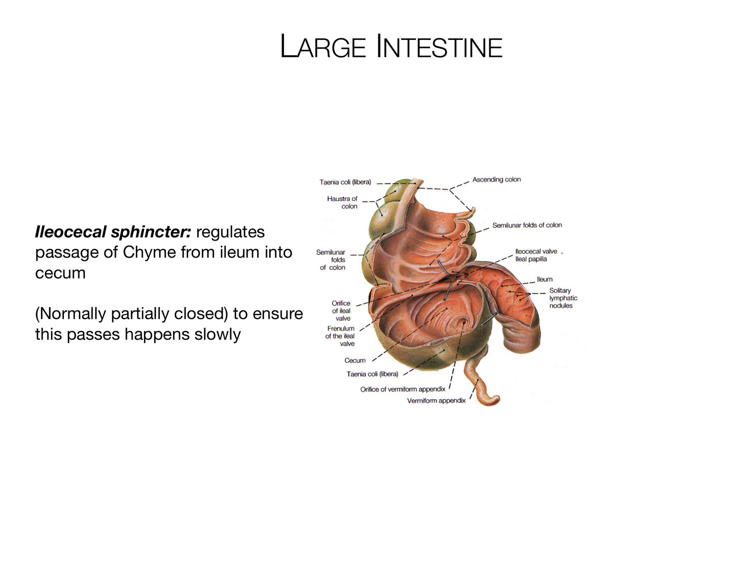
LARGE INTESTINE Ileocecal sphincter: regulates passage of Chyme from ileum into cecum (Normally partially closed) to ensure this passes happens slowly
Review the lecture
These slides

Open the full deck on your phone.
Practice from memory
Study-question guide

Comprehensive GI anatomy. Questions only.