BIO 304 · Human Anatomy & Physiology
Respiratory Anatomy & Mechanics
Respiratory System · Module 13
A reference for the Respiratory Anatomy video. The respiratory system has a conducting zone that moves air and a respiratory zone where gas exchange happens. Breathing is muscular work governed by pressure changes.
How to use this sheet Toggle the toolbar above. Notes prints the full reference for review. Study prints as a fill-in-the-blank worksheet , print it, then write each definition while you watch the video or read your book. Quiz me is on-screen typing practice; type the term, click Reveal to check yourself.
- Identify the structures of the upper and lower airway and the conducting versus respiratory zones.
- Explain how diaphragm and intercostal muscles produce inspiration and expiration.
- Define and distinguish lung volumes and capacities (TV, IRV, ERV, RV, VC, TLC).
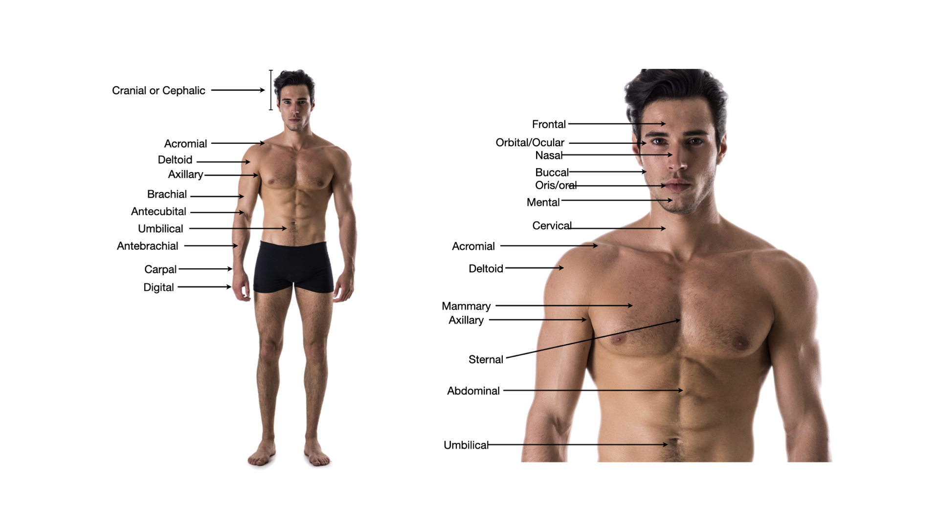
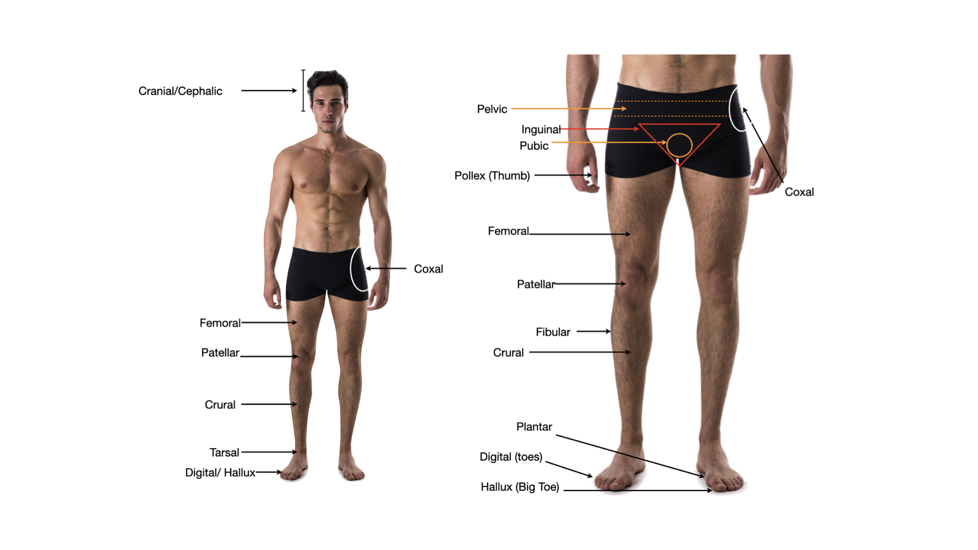
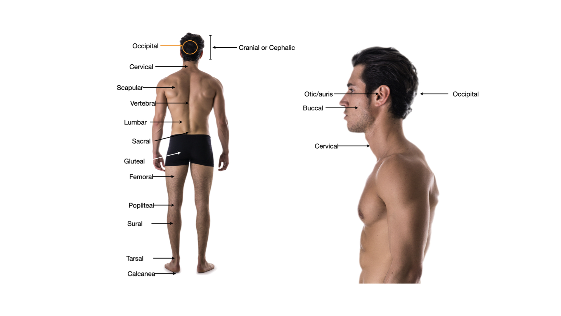
Click any image to enlarge.
Anatomy
Upper airway
- Nose & nasal cavitywarms, humidifies, filters; conchae increase surface area
- Pharynxnaso-, oro-, laryngo-; shared passage for air and food
- Larynxvoice box; epiglottis covers airway during swallowing
Lower airway: conducting zone
- TracheaC-shaped cartilage rings; ciliated columnar epithelium
- Main (primary) bronchiright + left; right is wider, shorter, more vertical
- Lobar (secondary) bronchi3 right, 2 left
- Segmental (tertiary) bronchifeed bronchopulmonary segments
- Bronchiolesno cartilage; smooth muscle controls diameter
- Terminal bronchioleslast conducting zone branch
Respiratory zone (gas exchange)
- Respiratory bronchiolesfirst scattered alveoli appear
- Alveolar ductslined with alveoli
- Alveolar sacsclusters of alveoli
- Alveoli~300 million; site of gas exchange; type I cells thin, type II make surfactant
Lungs & pleura
- Right lung3 lobes (sup, mid, inf)
- Left lung2 lobes (cardiac notch for heart)
- Visceral pleuracovers lung
- Parietal pleuralines thoracic wall and diaphragm
- Pleural cavitysmall fluid-filled space between pleurae; negative pressure
Mechanics & Volumes
Pressures
- Atmospheric (Patm)~760 mmHg at sea level
- Intrapulmonary (alveolar) pressurechanges with breathing; equals Patm at rest
- Intrapleural pressurealways sub-atmospheric (~-4 mmHg); keeps lungs expanded
- Transpulmonary pressurePalv minus Pip; keeps lung inflated
Inspiration (active)
- Diaphragm contractsflattens and moves down; main work
- External intercostalslift ribs up and out
- Thoracic volume increasespressure inside drops below atmospheric
- Air flows indown pressure gradient
Expiration (passive at rest)
- Diaphragm relaxesrises back to dome
- Lungs recoil elasticallyvolume decreases
- Pressure rises above Patmair flows out
- Forced expirationinternal intercostals + abdominal muscles add active force
Compliance & resistance
- Lung complianceease of expansion; reduced in fibrosis
- Airway resistancemostly bronchiolar; raised in asthma, COPD
- Surfactantlipoprotein from type II alveolar cells; lowers surface tension; prevents alveolar collapse
Lung volumes & capacities
- Tidal volume (TV)normal breath ~500 mL
- Inspiratory reserve volume (IRV)extra above TV with max inspiration
- Expiratory reserve volume (ERV)extra below TV with max expiration
- Residual volume (RV)cannot be exhaled; ~1200 mL; keeps alveoli open
- Vital capacity (VC)TV + IRV + ERV; max usable
- Total lung capacity (TLC)VC + RV
- FEV₁ / FVCfraction exhaled in first second; key in obstructive vs restrictive diagnosis
Step 3 . Retrieval check
Now explain it back, in your own words.
In 60 words or more, pull together what the video just taught you. Include the key concepts. This is the point where the learning actually sticks. After you submit, your spaced-recall cards for this topic unlock.
Zoomed image
Spaced recall unlocked
Nice work finishing the video.
Your spaced-recall cards for this topic are ready. Open the pre-work to activate them and start drilling.