BIO 304 · Human Anatomy & Physiology · Week 4 · Day 1

Muscle Anatomy

From how a muscle attaches and moves a joint, down to the connective tissue wrappers and the sarcomere inside.

Use the arrow keys, or the buttons below, to move through the slides.

What you will be able to do

Today’s objectives

  • 1State the functions of skeletal muscle and distinguish origin, insertion, and action.
  • 2Classify a muscle as agonist, antagonist, synergist, or fixator in a given movement.
  • 3Identify first, second, and third class levers and give a body example of each.
  • 4Trace the connective tissue hierarchy from epimysium down to a single myofibril.
  • 5Label the bands of the sarcomere and identify the T-tubules and sarcoplasmic reticulum.

Why we have it

What skeletal muscle does

Muscle is excitable, contractile, extensible, and elastic. Those four properties let it do five jobs.

  • MovementPulls on bones to move the body and its parts, and moves substances inside the body.
  • PostureConstant small adjustments hold you upright against gravity.
  • Joint stabilityMuscle tone braces joints that ligaments alone cannot hold.
  • HeatContraction releases heat; shivering is muscle making warmth on purpose.
  • GuardingSphincters and the body wall control openings and protect organs.

Three tissues, one job description

The three muscle types

All muscle is contractile, but the three types differ in structure, control, and where they work.

  • SkeletalStriated, multinucleate fibers; voluntary; attaches to bone and moves the skeleton. The focus of this unit.
  • CardiacStriated, short branching cells with one nucleus, joined by intercalated discs; involuntary and self-rhythmic; the heart wall only.
  • SmoothNot striated, spindle-shaped cells with one nucleus; involuntary; in the walls of hollow organs and vessels.

We build the contraction story on skeletal muscle, then compare how cardiac and smooth do it differently.
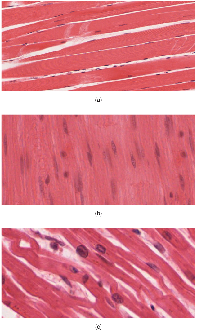
Micrographs comparing skeletal, smooth, and cardiac muscle tissue. Skeletal is striated with many nuclei, cardiac is striated with branching cells and intercalated discs, smooth is spindle-shaped with one central nucleus.
Histology of the three muscle types. OpenStax Anatomy & Physiology, CC BY 4.0
OpenStax cardiac muscle: histology micrograph and a diagram of intercalated discs with gap junctions and desmosomes.
Cardiac muscle. OpenStax Anatomy & Physiology, CC BY 4.0

How a muscle moves a bone

Origin, insertion, action

  • OriginThe attachment that stays relatively fixed when the muscle contracts.
  • InsertionThe attachment that moves. It is pulled toward the origin.
  • ActionThe movement produced, named at the joint crossed (for example, flexion of the elbow).
  • Belly and tendonsThe belly is the fleshy middle; tendons (or a flat aponeurosis) anchor each end to bone.

A muscle only pulls, it never pushes, and it must cross the joint it moves.

A muscle crossing a joint, animated to show flexion The origin stays fixed on the upper bone. As the muscle contracts, the insertion on the lower bone is pulled toward the origin and the joint flexes. The motion loops. Origin (fixed) belly Insertion (moves) action: flexion
The insertion is pulled toward the origin, flexing the joint.

Muscles work in teams

Agonist, antagonist, synergist, fixator

  • Agonist (prime mover)Mainly responsible for the movement. Biceps brachii in elbow flexion.
  • AntagonistOpposes the agonist and relaxes or lengthens to allow it. Triceps in elbow flexion.
  • SynergistAssists the agonist, adds force, or steadies the joint. Brachialis helping the biceps.
  • FixatorA synergist that stabilizes the origin so the movement is efficient. Rotator cuff steadying the shoulder.

Reverse the movement and the roles swap: in extension the triceps becomes the agonist.

Four muscle roles at the elbow During elbow flexion the biceps is the agonist, the triceps is the antagonist, the brachialis is a synergist assisting the biceps, and a shoulder muscle acts as a fixator that steadies the origin. Agonist biceps Antagonist triceps Synergist brachialis Fixator: steadies the shoulder
Four roles in one movement: agonist moves it, antagonist opposes it, synergist assists, fixator steadies the origin.

Bones, joints, and muscles make levers

The three lever classes

A lever is a rigid bar (bone) on a fulcrum (joint). Effort comes from the muscle; the load is the body part plus any weight. Memory hook: 1-2-3, the middle part is Fulcrum, then Load, then Effort.

First, second, and third class levers First class has the fulcrum in the middle, like a seesaw. Second class has the load in the middle, like a wheelbarrow. Third class has the effort in the middle, like tweezers, and is the most common in the body. Effort Load Fulcrum First class Fulcrum in the middle Like a seesaw Load Effort Fulcrum Second class Load in the middle Like a wheelbarrow Effort Load Fulcrum Third class Effort in the middle Like tweezers
Body examples: first class, nodding the head; second class, standing on tiptoe; third class, the biceps curl. Most muscles are third class: speed and range of motion over raw force.

The name tells you the anatomy

How muscles are named

Most names combine two or more of these clues, so reading the name often predicts location and action.

  • LocationWhere it sits. Tibialis anterior, intercostals.
  • ShapeIts outline. Deltoid (triangle), trapezius.
  • SizeRelative size. Gluteus maximus, minimus.
  • Fiber directionRectus (straight), oblique, transversus.
  • Number of headsBiceps (two), triceps (three), quadriceps (four).
  • AttachmentsOrigin then insertion. Sternocleidomastoid.
  • ActionWhat it does. Flexor, extensor, adductor.
OpenStax map of the major muscles of the body, anterior and posterior, superficial and deep.
Major muscles of the body. OpenStax Anatomy & Physiology, CC BY 4.0

Big to small

The connective tissue hierarchy

  • EpimysiumWraps the whole muscle. Dense irregular connective tissue.
  • PerimysiumWraps each fascicle, a bundle of muscle fibers.
  • EndomysiumWraps each fiber. The fiber membrane is the sarcolemma; its cytoplasm is the sarcoplasm.
  • Then insideMyofibrils, and within them the sarcomeres: the functional unit, Z-line to Z-line.
  • All three convergeEpimysium, perimysium, and endomysium merge at the ends to form the tendon.
Cross-section of a skeletal muscle The whole muscle is wrapped in epimysium. Inside are fascicles wrapped in perimysium, and each fascicle holds fibers wrapped in endomysium. Epimysium Perimysium Fiber +endomysium
Three wrappers nest from the whole muscle down to a single fiber.
OpenStax figure of the connective tissue layers of skeletal muscle: epimysium around the whole muscle, perimysium around a fascicle, and endomysium around each fiber, with the satellite cell, sarcolemma, and myofibril labeled.
Connective tissue layers of skeletal muscle. OpenStax Anatomy & Physiology, CC BY 4.0

Z-line to Z-line

Inside the sarcomere

  • A bandFull length of the thick filament. Width never changes during contraction.
  • I bandThin filaments only. Narrows during contraction.
  • H zoneThick filaments only. Narrows during contraction.
  • Z-line and M lineZ anchors thin filaments and bounds the sarcomere; M anchors thick filaments at the center.
Bands of the sarcomere The sarcomere runs from one Z-line to the next. The A band is the full length of the thick filaments. The I band has thin filaments only. The H zone is thick only, with the M line at the center. Z Z M line A band I band H zone
Thick filament is myosin (rust); thin filament is actin with troponin and tropomyosin (gold). Titin springs anchor the thick filament to the Z-line.
OpenStax figure of the thick and thin filaments in a sarcomere, labeling myosin, actin, troponin, and tropomyosin.
Thick and thin filaments. OpenStax Anatomy & Physiology, CC BY 4.0

Getting the signal in deep

Excitation machinery

These structures make sure the electrical signal reaches every myofibril at the same instant.

  • T-tubulesInvaginations of the sarcolemma that carry the action potential deep into the fiber, so contraction is uniform and not just at the surface.
  • Sarcoplasmic reticulumSmooth ER specialized for calcium storage. It releases Ca2+ the instant the T-tubule signal arrives, which triggers contraction.
  • The triadOne T-tubule flanked by two SR terminal cisternae. This is where the signal and the calcium meet.

Structure follows function: the T-tubule and SR sit side by side so the signal and the calcium meet in the same place at the same moment.

OpenStax figure of the triad: one T-tubule flanked by two sarcoplasmic reticulum terminal cisternae.
The triad. OpenStax Anatomy & Physiology, CC BY 4.0

Where the microanatomy shows up

Clinical tie-in

When a single structural protein fails, the whole fiber pays for it.

  • Duchenne muscular dystrophyA defect in dystrophin, the protein that anchors the contractile machinery to the sarcolemma. Without it, every contraction tears the fiber.
  • Statin-induced myopathyDisrupts the sarcoplasmic reticulum’s calcium handling, so contraction and relaxation go wrong.
  • The CK lab valueCreatine kinase rises when the sarcolemma is breached. Microanatomy is what fails first, even when the patient just feels weak.

Pull it together

Recap, then go practice

From the joint inward: attachments and levers set what a muscle does; the wrappers, sarcomere, and excitation machinery set how it does it. Print the workbook, label every band and lever yourself, then open your recall cards.

  • GrossFunctions; origin, insertion, action; agonist and antagonist; three lever classes.
  • MicroEpimysium, perimysium, endomysium, myofibril, sarcomere.
  • ExcitationT-tubules carry the signal in; the SR releases the calcium.

Next class: Muscle Physiology, how this anatomy actually contracts.

BIO 304 Human Anatomy & Physiology · American River College · Summer 2026 · Dr. Sharilyn Rennie