BIO 304 · Human Anatomy & Physiology · Week 4 · Day 2

Muscle Physiology

How yesterday’s anatomy actually contracts: the nerve signal, calcium, the cross-bridge cycle, and how whole muscles grade their force.

Use the arrow keys, or the buttons below, to move through the slides.

What you will be able to do

Today’s objectives

  • 1Trace the signal from the motor neuron across the neuromuscular junction to calcium release.
  • 2Describe the four steps of the cross-bridge cycle and how the muscle relaxes.
  • 3Explain motor units, the size principle, and the three fiber types.
  • 4Distinguish twitch, summation, and tetanus, and isotonic versus isometric contraction.
  • 5Compare the energy systems that fuel contraction and explain muscle fatigue.

One chain of events

From nerve signal to movement

Every voluntary contraction runs the same pathway. Keep this map in mind; the next slides zoom in on each link.

  1. A motor neuron fires and reaches the muscle fiber
  2. The neuromuscular junction passes the signal to the sarcolemma
  3. Excitation-contraction coupling releases calcium from the SR
  4. The cross-bridge cycle pulls the filaments and the sarcomere shortens
  5. Relaxation pumps calcium back and the fiber lengthens again

Where nerve meets muscle

The neuromuscular junction

A chemical synapse between a motor neuron and one muscle fiber. The transmitter is always acetylcholine.

  • Axon terminalThe nerve ending; holds vesicles of acetylcholine (ACh).
  • Synaptic cleftThe tiny gap the ACh diffuses across.
  • Motor end plateThe folded region of sarcolemma packed with nicotinic ACh receptors.
  • The hand-offAn action potential opens voltage-gated Ca2+ channels, vesicles fuse, ACh is released, binds receptors, and Na+ enters to start an end-plate potential.
  • AcetylcholinesteraseThe enzyme in the cleft that breaks down ACh so the signal stops cleanly.
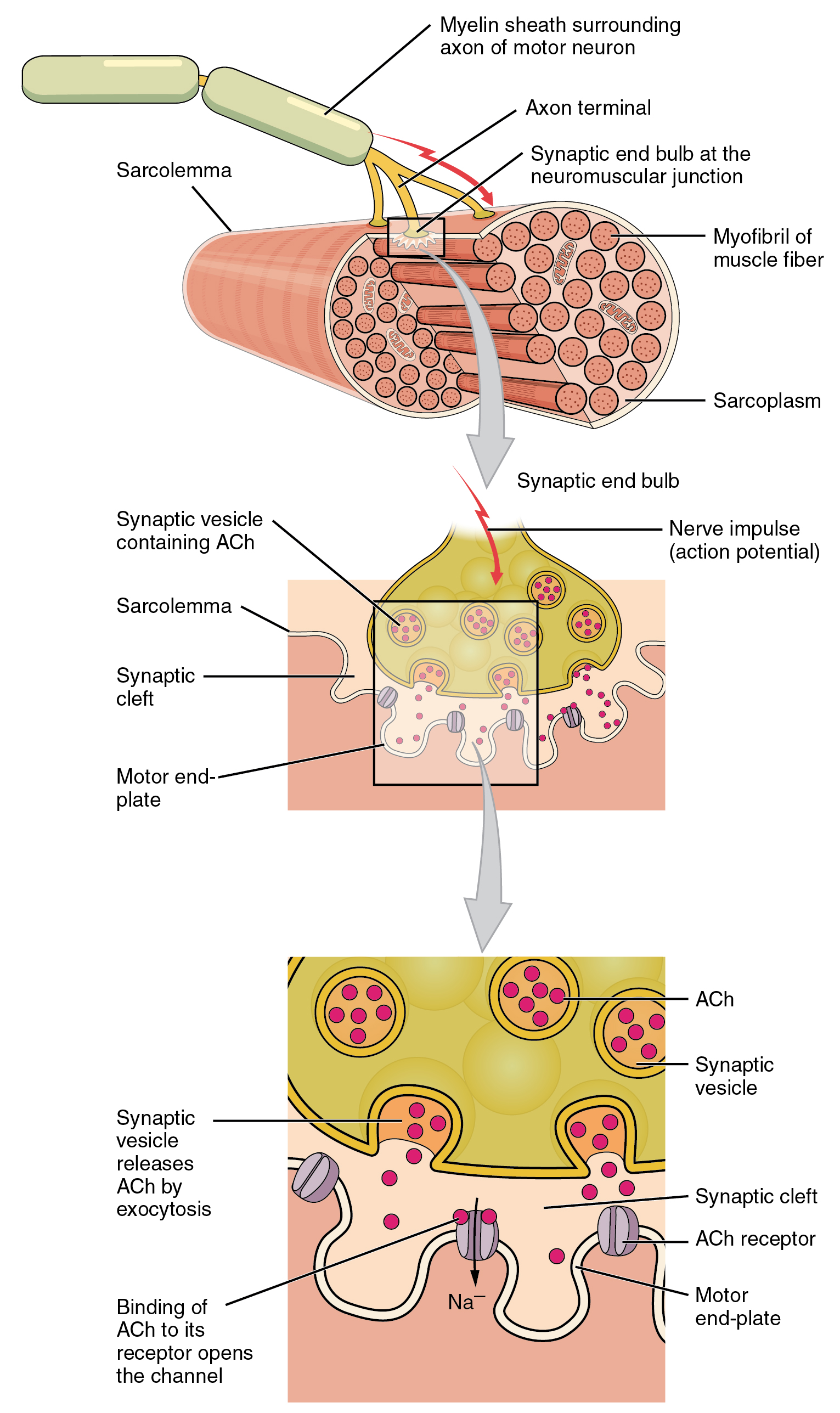
The neuromuscular junction with the motor neuron axon terminal, synaptic cleft, and the folded motor end plate bearing acetylcholine receptors.
The neuromuscular junction. OpenStax Anatomy & Physiology, CC BY 4.0

Turning a voltage into calcium

Excitation-contraction coupling

  1. The action potential sweeps along the sarcolemma and down the T-tubules
  2. Voltage sensors (DHP receptors) tug open the ryanodine receptors on the SR
  3. The SR floods the cytosol with Ca2+
  4. Ca2+ binds troponin, which pulls tropomyosin off the actin binding sites
  5. The myosin-binding sites are now exposed and contraction can begin

Calcium is the switch. No calcium, no exposed binding sites, no contraction.

Excitation-contraction coupling: the action potential travels down the T-tubule, the SR releases calcium, and calcium exposes the binding sites on actin.
Excitation-contraction coupling. OpenStax Anatomy & Physiology, CC BY 4.0

Four steps that repeat

The cross-bridge cycle

  1. Formation: an energized myosin head binds the exposed actin site
  2. Power stroke: the head pivots, pulling the thin filament toward the M line; ADP and Pi are released
  3. Detachment: a fresh ATP binds the myosin head, which lets go of actin
  4. Re-cocking: ATP splits to ADP and Pi, re-energizing the head to bind again

As long as calcium and ATP are present, the cycle repeats and the muscle keeps shortening.

The cross-bridge cycle in four stepsStep 1 the myosin head binds actin. Step 2 the power stroke pulls the thin filament. Step 3 ATP binds and the head detaches. Step 4 the head re-cocks, ready to bind again. 1 Bind 2 Power stroke ATP in 3 Detach ADP+Pi 4 Re-cock
Gold = thin filament (actin); rust = thick filament and myosin head. The head walks the thin filament toward the center.
The cross-bridge cycle: myosin binds actin, the power stroke pulls the thin filament, ATP binds and the head detaches, then re-cocks.
The cross-bridge cycle. OpenStax Anatomy & Physiology, CC BY 4.0

What shortening looks like

The sliding filament result

  • Thin filaments slideToward the M line from both ends of the sarcomere.
  • Z discs pulled inThe sarcomere gets shorter, and so does the whole fiber.
  • I band narrowsLess thin-only region.
  • H zone narrowsMore overlap of thick and thin.
  • A band unchangedThick filament length never changes, so the A band stays the same.

The filaments do not shrink. They slide past each other.
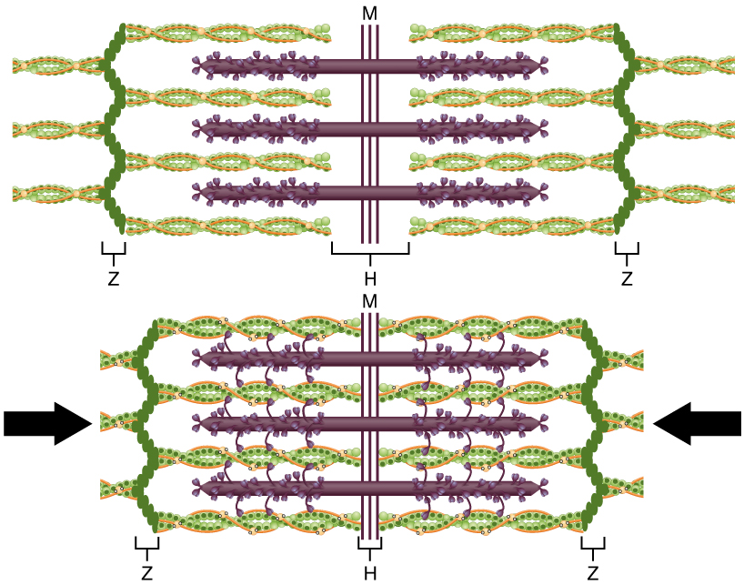
The sliding filament model showing a sarcomere relaxed and contracted, with the I band and H zone narrowing while the A band stays the same.
Sliding filament model. OpenStax Anatomy & Physiology, CC BY 4.0

Drag the slider or press play

Sliding filament, interactive

Turning it off

Relaxation, and when it cannot relax

  • Signal stopsThe motor neuron quiets and acetylcholinesterase clears the ACh.
  • SERCA pumpsActive transport (needs ATP) returns Ca2+ to the SR.
  • Tropomyosin recoversIt re-covers the actin sites, so cross-bridges can no longer form.
  • Rigor mortisAfter death there is no ATP, so myosin cannot release actin; the muscle locks until enzymes break the proteins down.
OpenStax figure of muscle relaxation: calcium is resorbed into the SR and the filaments slide back.
Relaxation. OpenStax Anatomy & Physiology, CC BY 4.0

Grading force, part 1

Motor units and recruitment

  • Motor unitOne motor neuron plus every muscle fiber it controls.
  • Small unitsFew fibers per neuron for fine control (eye muscles, about 3 to 5 fibers).
  • Large unitsThousands of fibers per neuron for gross force (calf, 1000+ fibers).
  • Size principleSmall, fatigue-resistant units fire first; larger ones are recruited as more force is needed.
  • All-or-noneA single fiber contracts fully or not at all; the muscle grades force by how many units it recruits.

Built for endurance or power

The three fiber types

  • Type I, slow oxidativeRed (lots of myoglobin and mitochondria), aerobic, fatigue-resistant. Posture and endurance.
  • Type IIa, fast oxidativeIntermediate; uses both aerobic and glycolytic fuel. Walking to jogging.
  • Type IIx, fast glycolyticWhite, anaerobic, powerful but fatigues fast. Sprinting and lifting.
  • Training noteEndurance training boosts Type I capacity; resistance training enlarges fast fibers.

Grading force, part 2

Twitch, summation, and tetanus

  • TwitchThe response to one action potential: latent, contraction, then relaxation phases.
  • Wave summationA second stimulus before relaxation adds onto the first, producing more force.
  • Unfused tetanusRapid stimulation with brief partial relaxation; force wobbles but climbs.
  • Fused tetanusStimulation so rapid there is no relaxation: smooth, maximal, sustained force.

Real movements use both recruitment (more units) and rate coding (faster firing) at once.

A myogram showing a single twitch, wave summation, unfused tetanus, and fused tetanus as stimulus frequency increases.
Twitch, summation, and tetanus. OpenStax Anatomy & Physiology, CC BY 4.0
OpenStax myogram of a single muscle twitch showing latent, contraction, and relaxation periods.
The muscle twitch. OpenStax Anatomy & Physiology, CC BY 4.0

When force is greatest, and kinds of contraction

Length-tension and contraction types

  • Optimal lengthMaximum filament overlap gives maximum force.
  • Too shortThick filaments hit the Z discs; force falls.
  • Too longToo little overlap to form bridges; force falls.
  • IsotonicMuscle changes length under a constant load (lifting a weight).
  • IsometricForce with no length change (holding a plank).
  • Concentric / eccentricConcentric shortens under load; eccentric lengthens under load and causes most soreness.
OpenStax length-tension graph: tension versus sarcomere length.
Length-tension relationship. OpenStax Anatomy & Physiology, CC BY 4.0
OpenStax figure of concentric, eccentric, and isometric contractions during a biceps curl.
Types of contraction. OpenStax Anatomy & Physiology, CC BY 4.0

Paying for contraction

Energy systems and fatigue

  • Stored ATPOn hand for about 2 seconds.
  • Creatine phosphateRegenerates ATP fast for about 10 to 15 seconds.
  • Anaerobic glycolysisAbout 30 to 60 seconds; makes ATP quickly but produces lactate.
  • Aerobic respirationMinutes to hours; needs oxygen, makes the most ATP.
  • Muscle fatigueA decline in force from ion imbalances, low energy, and reduced calcium release, not usually lactate alone.
  • Oxygen debtExtra oxygen used after exercise to restore ATP, creatine phosphate, and clear metabolites.
OpenStax figure of muscle energy systems: creatine phosphate, glycolysis, and aerobic respiration.
Muscle energy systems. OpenStax Anatomy & Physiology, CC BY 4.0

Same idea, different machinery

Cardiac and smooth muscle contraction

Both use actin, myosin, and calcium, but they are triggered and controlled differently from skeletal muscle.

  • Cardiac, the triggerSelf-rhythmic pacemaker cells start each beat; the signal spreads cell to cell through gap junctions in the intercalated discs.
  • Cardiac, the calciumCalcium entering from outside the cell triggers the SR to release more (calcium-induced calcium release).
  • Cardiac, no tetanusA long refractory period prevents sustained contraction, so the heart always relaxes and refills. The autonomic system and hormones adjust rate and force.
  • Smooth, the structureNo sarcomeres or striations; filaments anchor to dense bodies and twist the cell as they pull.
  • Smooth, the calciumCalcium binds calmodulin, which activates myosin light chain kinase to switch on myosin; slow, sustained, and energy efficient (latch state).
  • Smooth, the controlInvoluntary: driven by the autonomic system, hormones, and stretch. Single-unit sheets in the gut and vessels contract together.

Cardiac conduction and the full heartbeat get their own lecture in the cardiovascular week.
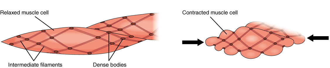
Smooth muscle contraction showing thin and thick filaments anchored to dense bodies, contracting the spindle-shaped cell.
Smooth muscle contraction. OpenStax Anatomy & Physiology, CC BY 4.0
OpenStax figure of smooth muscle: autonomic innervation and histology.
Smooth muscle. OpenStax Anatomy & Physiology, CC BY 4.0

When the physiology breaks

Clinical tie-in

  • Myasthenia gravisAntibodies destroy ACh receptors at the end plate, causing weakness that worsens with use.
  • Organophosphate poisoningBlocks acetylcholinesterase, so ACh builds up and muscles cannot relax (spasm, then paralysis).
  • Botulinum toxinBlocks ACh release, relaxing muscle; used clinically and cosmetically.
  • DOMSDelayed onset muscle soreness from the microdamage of eccentric contractions.

Pull it together

Recap, then go practice

Signal, calcium, cross-bridge, relaxation; then motor units, fiber types, and tetanus set how much force. Print the workbook, walk the cross-bridge cycle by hand, then open your recall cards.

Next class: Nervous Tissue, Organization, and Anatomy.

BIO 304 Human Anatomy & Physiology · American River College · Summer 2026 · Dr. Sharilyn Rennie