BIO 304 · Human Anatomy & Physiology
Bone Tissue & Bone Growth
Skeletal System · Module 5
A reference for the Bone Tissue video. Bone is a living, organ-system-active tissue. It builds, breaks down, and rebuilds throughout your life; it stores calcium and houses the marrow.
How to use this sheet Toggle the toolbar above. Notes prints the full reference for review. Study prints as a fill-in-the-blank worksheet , print it, then write each definition while you watch the video or read your book. Quiz me is on-screen typing practice; type the term, click Reveal to check yourself.
- Name the four bone cell types and the role of each in bone turnover.
- Distinguish compact from spongy bone by structure and function.
- Contrast intramembranous and endochondral ossification, and locate the epiphyseal plate.
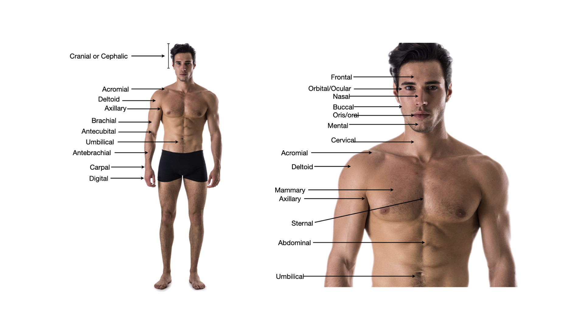
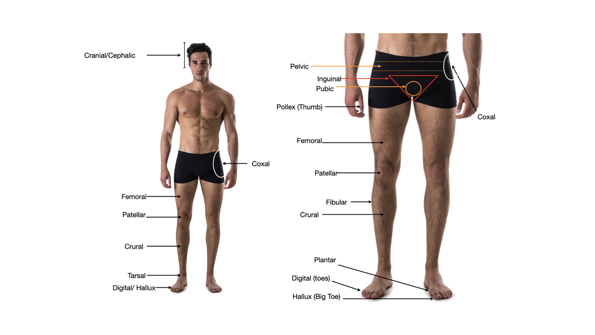
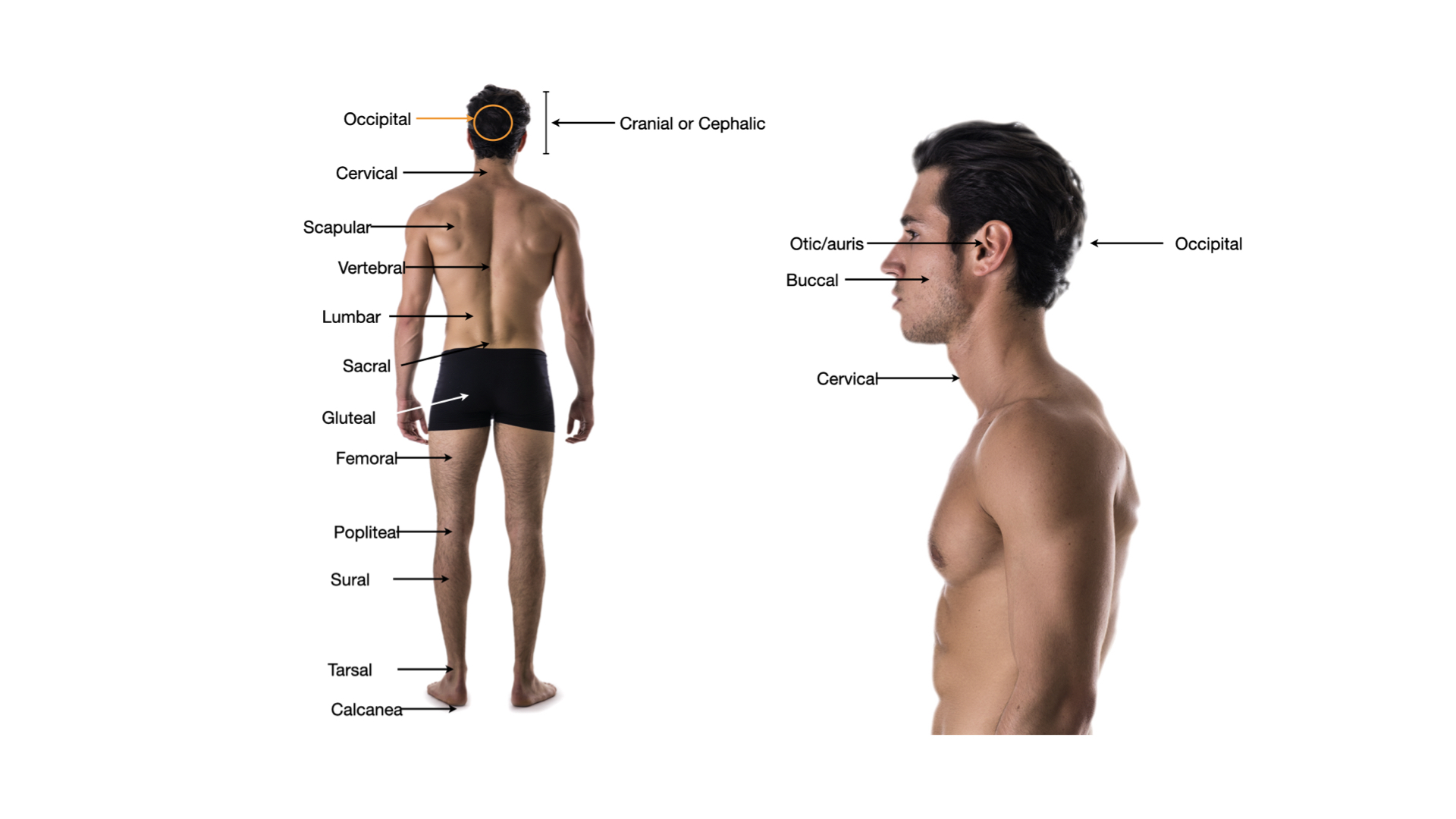
Click any image to enlarge.
Bone Cells & Matrix
Four bone cells
- Osteogenic (osteoprogenitor)stem cells in periosteum & endosteum; divide into osteoblasts
- Osteoblastbone builder; secretes osteoid (collagen + ground substance)
- Osteocytemature bone cell trapped in lacuna; maintains matrix
- Osteoclastmultinucleate giant cell; dissolves bone (resorption)
Matrix composition
- Organic (~33%)osteoid: collagen fibers + ground substance · tensile strength
- Inorganic (~67%)hydroxyapatite (calcium phosphate crystals) · compression strength
Compact bone
- Osteon (Haversian system)cylindrical unit of compact bone
- Central canalruns lengthwise; blood vessels and nerves
- Lamellaeconcentric rings of matrix
- Lacunaesmall spaces housing osteocytes
- Canaliculitiny channels connecting lacunae; nutrient diffusion
- Perforating (Volkmann) canalsrun perpendicular; connect central canals
Functions of the skeleton
- Support & shapeframework that holds up the body and cradles soft organs
- Protectionskull guards the brain; ribs guard heart and lungs; vertebrae guard the spinal cord
- Movement (leverage)bones act as levers that muscles pull on across joints
- Mineral storagereservoir for calcium and phosphate, released or stored on demand
- Blood cell formationred marrow makes red and white blood cells and platelets (hematopoiesis)
- Energy storageyellow marrow stores fat (triglycerides)
Bone shapes (classification)
- Longlonger than wide; humerus, femur, phalanges
- Shortcube-like; carpals, tarsals
- Flatthin, often curved; sternum, skull bones, ribs, scapula
- Irregularcomplex shape; vertebrae, hip bones
- Sesamoidforms within a tendon; patella
Long bone gross anatomy
- Diaphysisthe shaft; compact bone wall around the medullary cavity
- Epiphysiseach end; spongy bone under a thin compact shell
- Metaphysisregion between diaphysis and epiphysis; holds the growth plate
- Medullary cavityhollow center of the shaft; yellow marrow in adults
- Articular cartilagehyaline cartilage on the epiphysis tips; cushions joints
- Periosteumtough outer membrane; anchors tendons, carries vessels
- Endosteumthin membrane lining the medullary cavity
- Nutrient foramenopening where blood vessels enter the bone
Growth & Remodeling
Spongy (cancellous) bone
- Trabeculaelattice of bony struts; aligned along stress lines
- No osteonsosteocytes nourished by diffusion from marrow spaces
- Houses red marrowhematopoiesis — blood cell production
Ossification
- Intramembranousbone forms directly from mesenchyme · flat skull bones, clavicle
- Endochondralcartilage model is replaced by bone · most bones
- Primary ossification centerin diaphysis (shaft); forms first
- Secondary ossification centerin each epiphysis (end)
Growth in length & width
- Epiphyseal (growth) platecartilage between epiphysis and diaphysis; longitudinal growth
- Epiphyseal lineclosed growth plate — growth stopped (late teens/early 20s)
- Appositional growthosteoblasts add bone under periosteum; bone grows wider
Calcium homeostasis
- Parathyroid hormone (PTH)low blood Ca²⁺ → PTH ↑ → osteoclasts active → Ca²⁺ up
- Calcitoninhigh blood Ca²⁺ → calcitonin → deposits Ca²⁺ into bone
- Vitamin Denables Ca²⁺ absorption from the gut
Endochondral ossification, step by step
- 1. Cartilage modela hyaline cartilage version of the future bone forms
- 2. Bone collarosteoblasts build a bony collar around the shaft
- 3. Primary centercartilage in the shaft calcifies; osteoblasts lay down bone
- 4. Medullary cavityosteoclasts hollow out the center of the shaft
- 5. Secondary centersossification begins in each epiphysis
- 6. Cartilage remainsonly at articular surfaces and the epiphyseal plate
Epiphyseal plate: five zones
- Resting (reserve)anchors the plate to the epiphysis; quiet cartilage
- Proliferationchondrocytes divide and stack; the plate lengthens
- Hypertrophychondrocytes enlarge
- Calcificationmatrix calcifies; chondrocytes die
- Ossificationosteoblasts replace cartilage with bone
What controls bone growth
- Growth hormonemain driver of childhood length growth
- Thyroid hormonelets growth proceed at a normal pace
- Sex hormones (puberty)trigger the growth spurt, then close the plates
- Calcium & vitamin Dthe raw material and the means to absorb it
- Vitamin Cneeded to build the collagen in the matrix
- Mechanical stress (Wolff’s law)bone thickens where it is loaded, thins where it is not
Step 3 . Retrieval check
Now explain it back, in your own words.
In 60 words or more, pull together what the video just taught you. Include the key concepts. This is the point where the learning actually sticks. After you submit, your spaced-recall cards for this topic unlock.
Zoomed image
Spaced recall unlocked
Nice work finishing the video.
Your spaced-recall cards for this topic are ready. Open the pre-work to activate them and start drilling.