BIO 304 · Human Anatomy & Physiology
CNS Organization: Brain & Spinal Cord
Nervous System · Module 7
A reference for the CNS Organization video. The CNS is the brain plus the spinal cord, wrapped in meninges, bathed in CSF, and divided into gray matter (cell bodies) and white matter (myelinated tracts).
How to use this sheet Toggle the toolbar above. Notes prints the full reference for review. Study prints as a fill-in-the-blank worksheet , print it, then write each definition while you watch the video or read your book. Quiz me is on-screen typing practice; type the term, click Reveal to check yourself.
- Name the four major brain regions and the main functions of each.
- Identify the four lobes of the cerebrum and their primary roles.
- Describe the layers of the meninges and trace CSF flow through the ventricles.
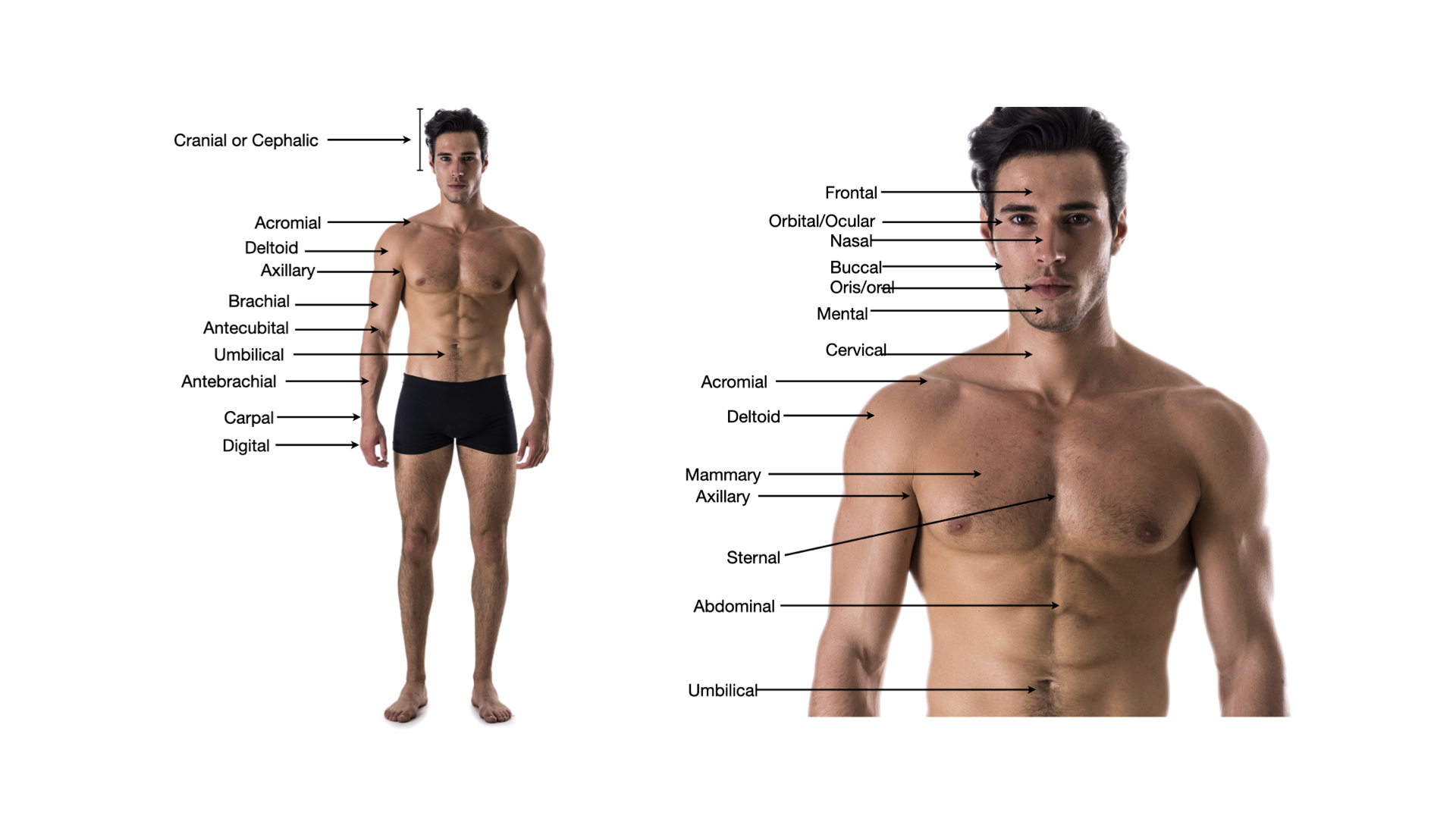
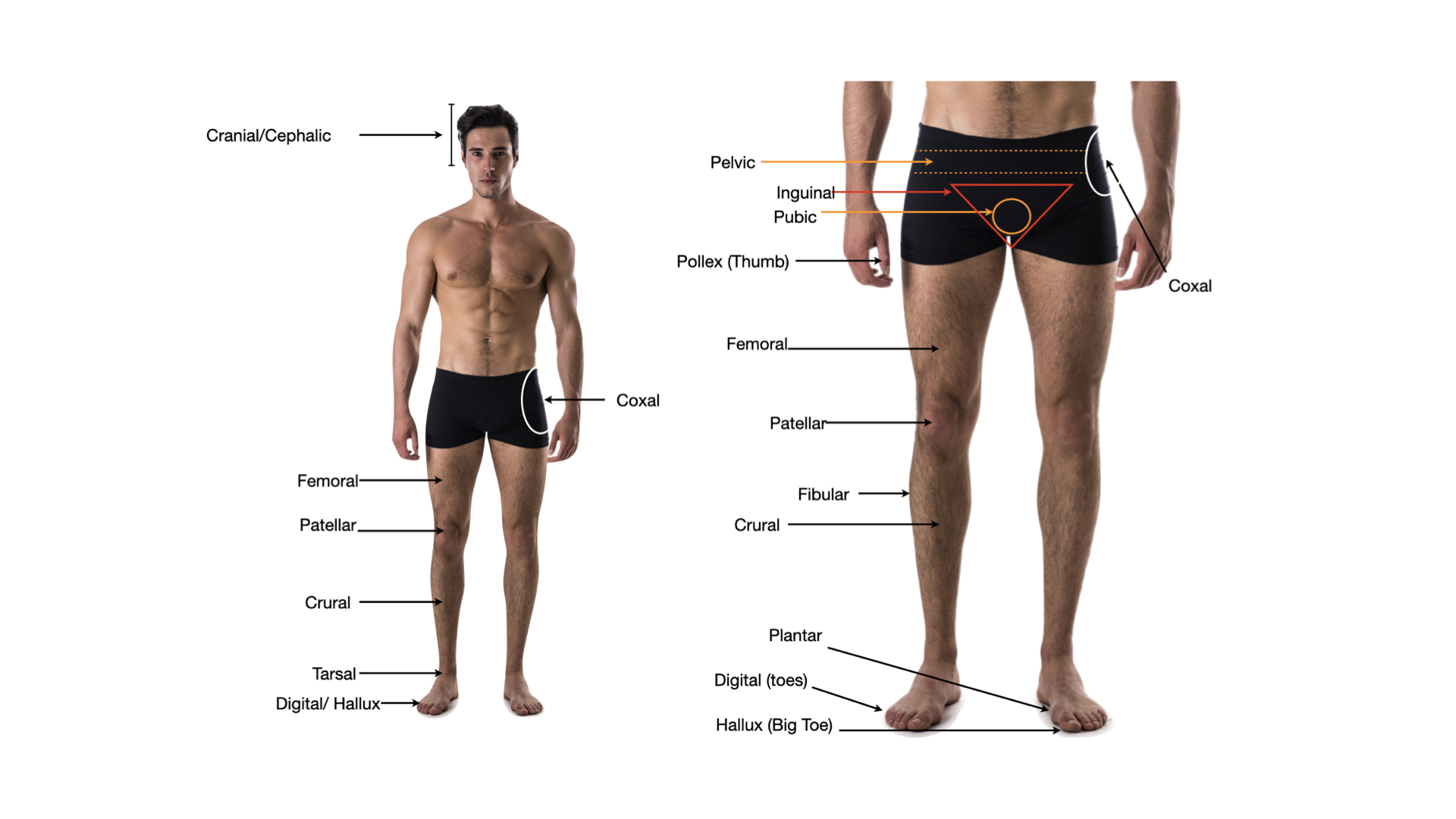
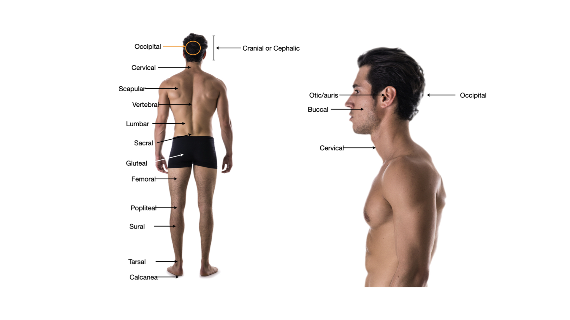
Click any image to enlarge.
Brain
Four major regions
- Cerebrumlargest; cognition, sensation, voluntary movement
- Diencephalonthalamus + hypothalamus + epithalamus
- Brainstemmidbrain + pons + medulla; vital reflexes
- Cerebellumcoordination, balance, motor learning
Cerebral lobes
- Frontalvoluntary motor (precentral gyrus), executive function, personality, Broca speech
- Parietalsomatosensation (postcentral gyrus), spatial orientation
- Temporalhearing, language comprehension (Wernicke), memory
- Occipitalvision
- Insuladeep; taste, interoception
Diencephalon
- Thalamussensory relay station (except smell)
- Hypothalamushomeostasis: temp, hunger, thirst, hormones via pituitary
- Pineal gland (epithalamus)melatonin; circadian rhythm
Brainstem
- Midbrainvisual and auditory reflexes; substantia nigra
- Ponsbridge between cerebrum and cerebellum; respiratory control
- Medullacardiac, vasomotor, respiratory centers; decussation of pyramids
Spinal Cord & Coverings
Spinal cord
- Extendsfrom medulla to L1/L2 in adults
- 31 pairsof spinal nerves exit through intervertebral foramina
- Cervical & lumbar enlargementswhere limb-innervating nerves arise
- Cauda equinalumbar/sacral nerve roots below L1/L2
- Conus medullaristapered end of cord (around L1)
Cross-section
- Gray matter (H-shape)cell bodies; central
- Dorsal hornsensory input from afferent neurons
- Ventral hornmotor neuron cell bodies
- Lateral horn (T1-L2)sympathetic preganglionic neurons
- White matter columnsascending/descending tracts
- Central canalCSF down the middle
Meninges (outside → in)
- Dura matertough outer layer
- Arachnoid materweb-like middle; subarachnoid space holds CSF
- Pia materthin, follows every contour of brain
CSF
- Produced bychoroid plexus in the ventricles
- Flowlateral → 3rd ventricle → cerebral aqueduct → 4th ventricle → subarachnoid space → arachnoid villi → venous blood
- Functionscushion, buoyancy, chemical stability
Blood-brain barrier
- Built bytight junctions between brain capillary endothelial cells + astrocyte feet
- Lets throughlipid-soluble molecules, glucose (via transporter)
- Excludesmost drugs and toxins; protects neurons
Step 3 . Retrieval check
Now explain it back, in your own words.
In 60 words or more, pull together what the video just taught you. Include the key concepts. This is the point where the learning actually sticks. After you submit, your spaced-recall cards for this topic unlock.
Zoomed image
Spaced recall unlocked
Nice work finishing the video.
Your spaced-recall cards for this topic are ready. Open the pre-work to activate them and start drilling.