BIO 304 · Human Anatomy & Physiology
Epithelial Tissue Classification
Tissues · Module 3
A reference for the Epithelial Tissue video. Epithelium covers, lines, and secretes. Every epithelium gets named twice: by shape of cell, and by how many layers thick.
How to use this sheet Toggle the toolbar above. Notes prints the full reference for review. Study prints as a fill-in-the-blank worksheet , print it, then write each definition while you watch the video or read your book. Quiz me is on-screen typing practice; type the term, click Reveal to check yourself.
- Classify any epithelium by cell shape (squamous, cuboidal, columnar) and number of layers (simple, stratified, pseudostratified).
- Match each epithelium to a representative location and primary function.
- Distinguish exocrine from endocrine glands and name the three modes of exocrine secretion.
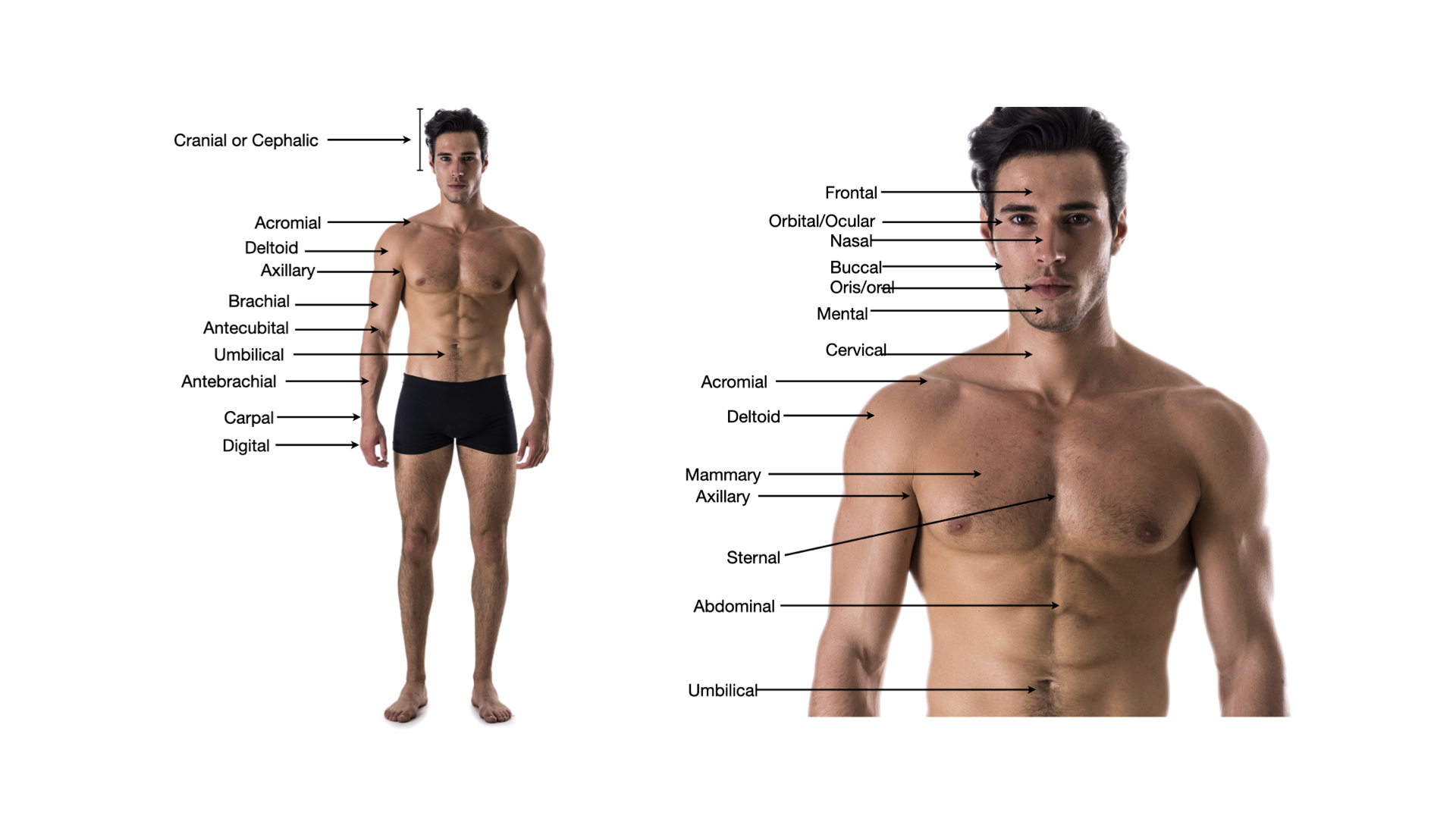
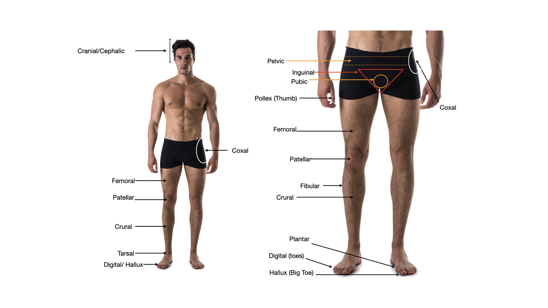
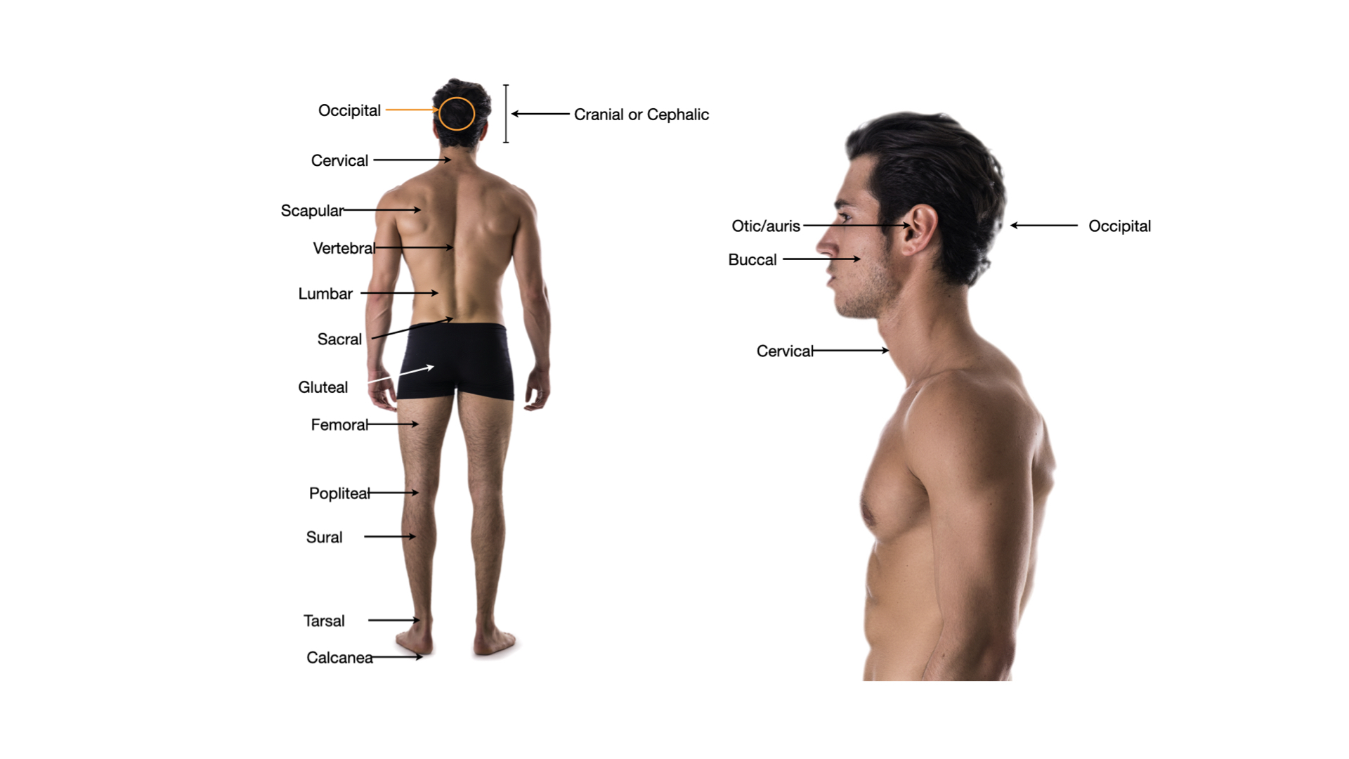
Click any image to enlarge.
By Shape & Layers
Cell shapes
- Squamousflat, scale-like cells; thin barrier for diffusion
- Cuboidalcube-shaped; secretion and absorption
- Columnartall column-shaped; absorption, secretion, sometimes cilia
Layering
- Simpleone cell layer; thin enough for transport
- Stratifiedmultiple cell layers; protection against abrasion
- Pseudostratifiedone layer but nuclei at different heights, looks layered
- Transitionalchanges shape with stretch; only in urinary tract
Common types & where
- Simple squamousalveoli, capillary endothelium · gas/fluid exchange
- Simple cuboidalkidney tubules, gland ducts
- Simple columnarGI tract lining · absorption + mucus
- Pseudostratified ciliated columnarupper airway · mucus + cilia escalator
- Stratified squamousskin epidermis, esophagus, vagina · abrasion
- Transitional (urothelium)bladder, ureters · stretch without leaking
Glandular Epithelium
Gland types
- Exocrinesecretes through a duct to a surface (sweat, salivary, mammary)
- Endocrineductless; secretes hormones into bloodstream (thyroid, adrenal)
- Unicellular glanda single secretory cell (goblet cell → mucus)
- Multicellular glandorganized acini and ducts (pancreas, salivary)
Modes of secretion
- Merocrineexocytosis only; cell intact (sweat glands, salivary, pancreas)
- Apocrineapical portion of cell pinches off (mammary, some sweat)
- Holocrinewhole cell ruptures; replaced (sebaceous glands)
Universal features
- Apical surfacetop, faces lumen or surface; may have cilia or microvilli
- Basal surfacebottom, sits on basement membrane
- Avascularno blood vessels in the tissue; nourished by diffusion
- High regenerationcontinuous turnover (gut lining renews every 3-5 days)
Step 3 . Retrieval check
Now explain it back, in your own words.
In 60 words or more, pull together what the video just taught you. Include the key concepts. This is the point where the learning actually sticks. After you submit, your spaced-recall cards for this topic unlock.
Zoomed image
Spaced recall unlocked
Nice work finishing the video.
Your spaced-recall cards for this topic are ready. Open the pre-work to activate them and start drilling.