BIO 304 · Human Anatomy & Physiology
Joints & Body Movements
Skeletal System · Module 5
A reference for the Joints & Movements video. Joints are everywhere two bones meet. They are classified by what tissue holds them together (structural) or how freely they move (functional).
How to use this sheet Toggle the toolbar above. Notes prints the full reference for review. Study prints as a fill-in-the-blank worksheet , print it, then write each definition while you watch the video or read your book. Quiz me is on-screen typing practice; type the term, click Reveal to check yourself.
- Classify a joint by structural type (fibrous, cartilaginous, synovial) and functional type (synarthrosis, amphiarthrosis, diarthrosis).
- Identify the six types of synovial joints and give an example of each.
- Define and demonstrate the major body movements (flexion/extension, abduction/adduction, rotation, circumduction, etc.).
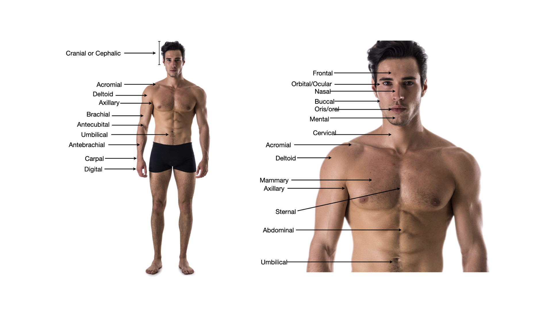
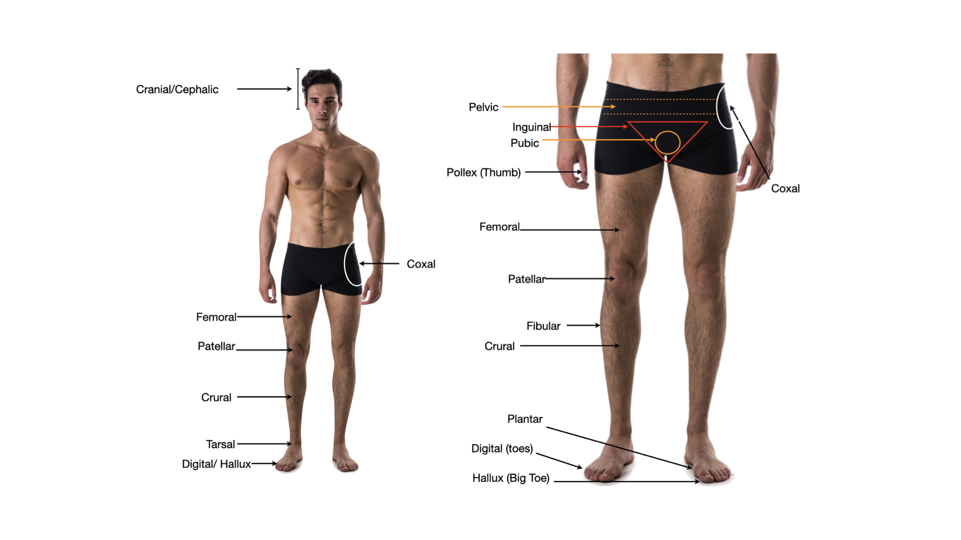
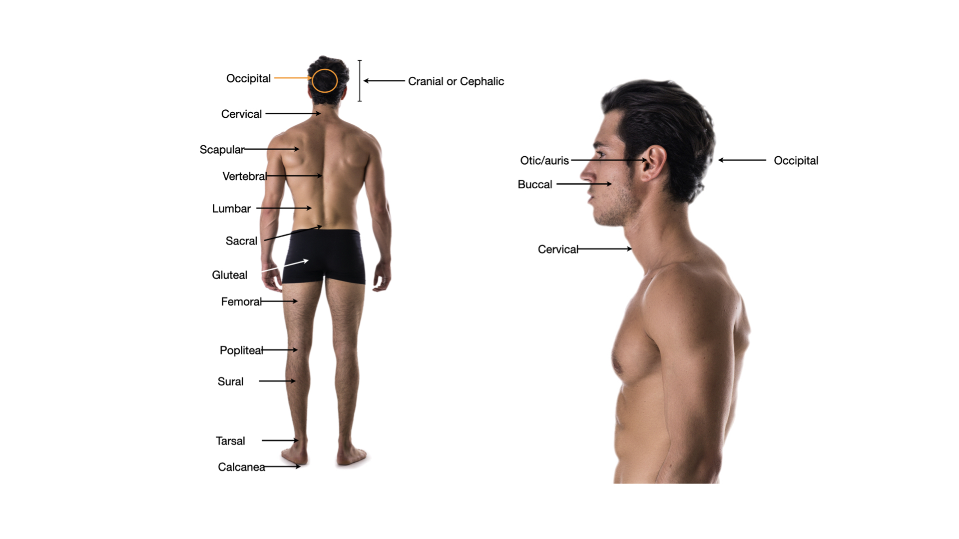
Click any image to enlarge.
Joint Classification
Structural (by tissue)
- Fibrousbones held by dense connective tissue; little or no movement
- Cartilaginousbones held by cartilage; little movement
- Synovialfluid-filled joint cavity; freely movable
Functional (by movement)
- Synarthrosisimmovable (sutures of the skull)
- Amphiarthrosisslightly movable (intervertebral discs, pubic symphysis)
- Diarthrosisfreely movable (all synovial joints)
Synovial joint features
- Articular cartilagehyaline cartilage covering bone ends; reduces friction
- Joint (synovial) cavityspace between articulating bones
- Synovial membraneinner lining; makes synovial fluid
- Synovial fluidlubricant + nutrient supply for cartilage
- Joint capsulefibrous outer envelope; reinforced by ligaments
- Bursaefluid sacs that cushion structures around the joint
Synovial Subtypes & Movements
Six synovial joint types
- Plane (gliding)flat surfaces glide; intercarpal, intertarsal
- Hingeone axis · elbow, knee, interphalangeal
- Pivotrotation around one axis · atlantoaxial, proximal radioulnar
- Condyloid (ellipsoid)two axes · wrist, metacarpophalangeal
- Saddletwo axes, greater range than condyloid · thumb (carpometacarpal)
- Ball-and-socketthree axes · shoulder, hip
Angular movements
- Flexiondecreases angle (bending the elbow)
- Extensionincreases angle (straightening the elbow)
- Hyperextensionbeyond anatomical position
- Abductionaway from the midline
- Adductiontoward the midline
- Circumductioncone-shaped movement combining all four
Rotational & special
- Rotationturning around long axis
- Supination / Pronationpalm up / palm down at the forearm
- Inversion / Eversionsole turns medially / laterally
- Dorsiflexion / Plantar flexionfoot up / foot down
- Oppositionthumb tip to fingertip
- Elevation / Depressionshoulders up / down (shrug)
Step 3 . Retrieval check
Now explain it back, in your own words.
In 60 words or more, pull together what the video just taught you. Include the key concepts. This is the point where the learning actually sticks. After you submit, your spaced-recall cards for this topic unlock.
Zoomed image
Spaced recall unlocked
Nice work finishing the video.
Your spaced-recall cards for this topic are ready. Open the pre-work to activate them and start drilling.